Täällä on ensin niitä "sellaisia" "tutkimuksia", joissa on tuossa huijattu tai ainakin menty muuten vaan metsään:
www.nature.com/neuro/journal/v11/n5/full/nn.2112.html
www.rifters.com/real/articles/NatureNeuroScience_Soon_et_al.pdf
homepages.abdn.ac.uk/a.hunt/pages/dept/L4Option/Haggard2005.pdf
web.gc.cuny.edu/cogsci/private/wegner-trick.pdf
Täällä on EU:n tutkimuslaitoksen tyylinäyte tässä asiasaa vuodelta 2008, jolloin Fields astui kuvioihin vislaamaan hölynpölyä poikki:
www.nature.com/news/2008/080411/full/news.2008.751.html
" NATURE 11.04.2008
Brain makes decisions before you even know it
Brain activity predicts decisions before they are consciously made. Kerri Smith
Your brain makes up its mind up to ten seconds before you realize it,according to re-searchers. By looking at brain activity while making a decision,the researchers could predict what choice people would make before they themselves were even aware of having made a decision.
The work calls into question the ‘consciousness’ of our decisions and may even challenge ideas about how ‘free’ we are to make a choice at a particular point in time.
“We think our decisions are conscious, but these data show that consciousness is just the tip of the iceberg", says John-Dylan Haynes, a neuroscientist at the Max Planck Insstitute for Human Cognitive and Brain Sciences in Leipzig, Germany, who led the study.
“ The results are quite dramatic", says Frank Tong,a neuroscientist at Vanderbilt Uni-versity in Nashville, Tennessee. Ten seconds is "a lifetime” in terms of brain activity, he adds.
On the button
Haynes and his colleagues imaged the brains of 14 volunteers while they performed a decision-making task.The volunteers were asked to press one of two buttons when they felt the urge to. Each button was operated by a different hand.At the same time, a stream of letters were presented on a screen at half-second intervals, and the vo-lunteers had to remember which letter was showing when they decided to press their button.
When the researchers analysed the data,the earliest signal the team could pick up started seven seconds before the volunteers reported having made their decision. Because of there is a delay of a few seconds in the imaging, this means that the brain activity could have begun as much as ten seconds before the conscious deci-sion. The signal came from a region called the frontopolar cortex, at the front of the brain, immediately behind the forehead.
This area may well be the brain region where decisions are initiated, says Haynes, who reports the results online in Nature Neuroscience 1.
The next step is to speed up the data analysis to allow the team to predict people's choices as their brains are making them.
Mind over matter
The results build on some well-known work on free will done in the 1980s by the late neurophysiologist Benjamin Libet, then at the University of California, San Francisco. Libet used a similar experimental set-up to Haynes, but with just one button and measuring electrical activity in his subjects' brains. He found that the regions respon-sible for movement reacted a few hundred milliseconds before a conscious decision was made.
But Libet's study has been criticized in the intervening decades for its method of measuring time, and because the brain response might merely have been a general preparation for movement,rather than activity relating to a specific decision. Haynes and his team improved the method by asking people to choose between two alterna-tives - left and right. Because moving the left and right hands generates distinct brain signals, the researchers could show that activity genuinely reflected one of the two decisions.
But the experiment could limit how ‘free’ people’s choices really are,says Chris Frith, who studies consciousness and higher brain function at University College London. Although subjects are free to choose when and which button to press, the experi-mental set-up restricts them to only these actions and nothing more, he says. “The subjects hand over their freedom to the experimenter when they agree to enter the scanner," he says.
What might this mean,then,for the nebulous concept of free will? If choices really are being made several seconds ahead of awareness, “there’s not much space for free will to operate”, Haynes says.
But results aren't enough to convince Frith that free will is an illusion. “We already know our decisions can be unconsciously primed,” he says.The brain activity could be part of this priming, as opposed to the decision process, he adds.
Part of the problem is defining what we mean by ‘free will’. But results such as these might help us settle on a definition. It is likely that “neuroscience will alter what we mean by free will”, says Tong. ”
Kaikki tekijät ovat tunnettuja haistpaskantiteilijöitä. Lisää samaa:
https://www.wired.com/2008/04/mind-decision/
Brandon Keim Science
Brain Scanners Can See Your Decisions Before You Make Them

You may think you decided to read this story - but in fact, your brain made the decision long before you knew about it.
This article has been reproduced in a new format and may be missing content or contain faulty links. Contact [email protected] to report an issue.
In a study published Sunday in Nature Neuroscience, researchers using brain scan-ners could predict people's decisions seven seconds before the test subjects were even aware of making them.
The decision studied - whether to hit a button with one's left or right hand - may not be representative of complicated choices that are more integrally tied to our sense of self-direction. Regardless, the findings raise profound questions about the nature of self and autonomy: How free is our will? Is conscious choice just an illusion?
"Your decisions are strongly prepared by brain activity. By the time consciousness kicks in, most of the work has already been done," said study co-author John-Dylan Haynes, a Max Planck Institute neuroscientist.
Haynes updated a classic experiment by the late Benjamin Libet, who showed that a brain region involved in coordinating motor activity fired a fraction of a second before test subjects chose to push a button. Later studies supported Libet's theory that sub-conscious activity preceded and determined conscious choice - but none found such a vast gap between a decision and the experience of making it as Haynes' study has .
In the seven seconds before Haynes' test subjects chose to push a button, activity shifted in their frontopolar cortex, a brain region associated with high-level planning. Soon afterwards,activity moved to the parietal cortex,a region of sensory integration. Haynes' team monitored these shifting neural patterns using a functional MRI machine.
Taken together,the patterns consistently predicted whether test subjects eventually pushed a button with their left or right hand - a choice that, to them, felt like the out-come of conscious deliberation. For those accustomed to thinking of them-selves as having free will, the implications are far more unsettling than learning about the physiological basis of other brain functions.
Caveats remain,holding open the door for free will. For instance,the experiment may not reflect the mental dynamics of other, more complicated decisions.
"Real-life decisions - am I going to buy this house or that one, take this job or that - aren't decisions that we can implement very well in our brain scanners",said Haynes.
Also, the predictions were not completely accurate. Maybe free will enters at the last moment, allowing a person to override an unpalatable subconscious decision.
"We can't rule out that there's a free will that kicks in at this late point", said Haynes, who intends to study this phenomenon next. "But I don't think it's plausible."
That implausibility doesn't disturb Haynes.
"It's not like you're a machine. Your brain activity is the physiological substance in which your personality and wishes and desires operate," he said.
The unease people feel at the potential unreality of free will,said National Institutes of Health neuroscientist Mark Hallett, originates in a misconception of self as separate from the brain.
"That's the same notion as the mind being separate from the body - and I don't think anyone really believes that," said Hallett. "A different way of thinking about it is that your consciousness is only aware of some of the things your brain is doing."
Hallett doubts that free will exists as a separate, independent force.
"If it is, we haven't put our finger on it," he said. "But we're happy to keep looking."
Ja sitten kunnon tukimusta:
www.nature.com/neuro/journal/v15/n4/full/nn.3045.html?WT.ec_id=NEURO-201204#/ref1
" Nature Neuroscience | Review
Plasticity in gray and white: neuroimaging changes in brain structure during learning
Robert J Zatorre, R Douglas Fields & Heidi Johansen-Berg
Nature Neuroscience 15, 528–536(2012) doi:10.1038/nn.3045 0Published online 18 March 2012
Abstract
Human brain imaging has identified structural changes in gray and white matter that occur with learning. However, ascribing imaging measures to underlying cellular and molecular events is challenging. Here we review human neuroimaging findings of structural plasticity and then discuss cellular and molecular level changes that could underlie observed imaging effects. Greater dialog between researchers in these different fields would help to facilitate cross-talk between cellular and systems level explanations of how learning sculpts brain structure.
The brain is the source of behavior,but in turn it is modified by the behaviors it produ- ces.This dynamic loop between brain structure and brain function is at the root of the neural basis of cognition, learning and plasticity. The concept that brain structure can be modified by experience is not new, but it has proven difficult to address experi-mentally. Recent developments in structural brain imaging techniques (Figure 1, Box 1), particularly magnetic resonance imaging (MRI), are now propelling such studies to the forefront of human cognitive neuroscience.
A connection between brain function and brain anatomy might be expected because neural information processing depends on the size,configuration and arrangement of individual neurons; on the number and type of local synaptic connections they make; on the way that they are interconnected to distant neuronal populations; and on pro-perties of non-neuronal cells, such as glia. Neuroimaging evidence, reviewed below, shows both differences in structural features among individuals and the relevant functions that these structures subserve, and changes in structural features when long-term neural activity patterns are changed by experience.
However, existing neuroimaging techniques cannot directly inform us about the un-derlying cellular events mediating the observed effects. Moreover,phenomena visible with MRI are likely never the result of a single process happening independently, but probably involve many coordinated structural changes involving various cell types.
Conversely,neuroimaging techniques offer certain advantages,as they can be repea- tedly performed in the same individual and provide whole-brain measures of brain structure and function. Contemporary neural models of cognition stress the idea of interacting functional networks; it is therefore logical to search for network-level pat-terns in anatomical structures as well. Recent studies examining interregional corre-lations of cortical thickness reveal that gray matter anatomical net-works parallel functional organizational patterns1,that they are modified during development 2 and that they are sensitive to training 3. The ability provided by macrostructural imaging methods to understand both function and anatomy in terms of regional interactions is likely to grow in importance and can also help to create hypotheses to which cellular and molecular probes can be applied.
Here we consider findings that have emerged from the human anatomical neuroima-ging literature,discuss the questions raised by them and propose some possible mic- rostructural mechanisms that could underlie the observed macrostructural findings.
Tiivistelmä
Ihmisen aivojen kuvantaminen on tunnistanut harmaan ja valkean aineen rakenteelli-sia muutoksia, joita tapahtuu oppimisen yhteydessä. Kuvantamismittausten yhdistä-minen taustalla oleviin solu- ja molekyylitason tapahtumiin on kuitenkin haastavaa.
Tässä tarkastelemme ihmisen neurokuvantamisen löydöksiä rakenteellisesta plasti-suudesta ja keskustelemme sitten solu- ja molekyylitason muutoksista, jotka voisivat olla havaittujen kuvantamisvaikutusten taustalla. Näiden eri alojen tutkijoiden välinen laajempi vuoropuhelu auttaisi helpottamaan vuorovaikutusta solu- ja järjestelmäta-son selitysten välillä siitä, miten oppiminen muokkaa aivojen rakennetta.
Aivot ovat käyttäytymisen lähde, mutta niitä puolestaan muokkaavat niiden tuottamat käyttäytymismallit. Tämä dynaaminen silmukka aivojen rakenteen ja toiminnan välillä on kognition, oppimisen ja plastisuuden hermostollisen perustan ytimessä. Ajatus sii-tä,että aivojen rakennetta voidaan muokata kokemuksen avulla,ei ole uusi,mutta sen käsitteleminen kokeellisesti on osoittautunut vaikeaksi. Viimeaikaiset kehitysaskeleet rakenteellisissa aivokuvantamistekniikoissa (kuva 1 laatikko 1), erityisesti magneetti-kuvauksessa (MRI), nostavat nyt tällaiset tutkimukset ihmisen kognitiivisen neurotieteen eturintamaan.
Yhteyttä aivotoiminnan ja aivojen anatomian välillä voidaan odottaa, koska hermos-tollinen tiedonkäsittely riippuu yksittäisten hermosolujen koosta, kokoonpanosta ja järjestyksestä; niiden muodostamien paikallisten synaptisten yhteyksien lukumääräs-tä ja tyypistä; tavasta, jolla ne ovat yhteydessä etäisiin hermosolupopulaatioihin; ja ei-neuroni-solujen kuten gliasolujen, ominaisuuksista. Alla tarkastellut neurokuvanta-mistutkimukset osoittavat sekä yksilöiden välisiä eroja rakenteellisissa ominaisuuk-sissa että näiden rakenteiden suorittamissa merkityksellisissä toiminnoissa, että muutoksia rakenteellisissa ominaisuuksissa, kun kokemus muuttaa pitkäaikaisia hermostollisia toimintamalleja.
Nykyiset neurokuvantamistekniikat eivät kuitenkaan voi suoraan antaa tietoa havait-tujen vaikutusten taustalla olevista solutapahtumista. Lisäksi magneettikuvauksella näkyvät ilmiöt eivät todennäköisesti ole koskaan yksittäisen itsenäisen prosessin tu-losta, vaan niihin liittyy todennäköisesti monia koordinoituja rakenteellisia muutoksia, joihin osallistuu eri solutyyppejä.
Kääntäen, neurokuvantamistekniikat tarjoavat tiettyjä etuja, koska niitä voidaan suo-rittaa toistuvasti samalla yksilöllä ja ne tarjoavat koko aivojen mittauksia aivojen ra-kenteesta ja toiminnasta. Nykyaikaiset kognition hermostolliset mallit korostavat vuo-rovaikutteisten toiminnallisten verkostojen ajatusta; siksi on loogista etsiä verkostota-son malleja myös anatomisista rakenteista. Viimeaikaiset tutkimukset, joissa on tar-kasteltu aivokuoren paksuusalueiden välisiä korrelaatioita, paljastavat, että harmaan aineen anatomiset verkostot ovat samansuuntaisia toiminnallisten organisaatiomal-lien kanssa 1, että ne muuttuvat kehityksen aikana 2 ja että ne ovat herkkiä harjoitte-lulle 3. Makrorakenteellisten kuvantamismenetelmien tarjoama kyky ymmärtää sekä toimintaa että anatomiaa alueellisten vuorovaikutusten kautta todennäköisesti kasvaa ja voi myös auttaa luomaan hypoteeseja, joihin solu- ja molekyylimittauksia voidaan soveltaa.
Tässä tarkastelemme ihmisen anatomisen neurokuvantamisen kirjallisuudesta esiin tulleita havaintoja, keskustelemme niiden herättämistä kysymyksistä ja ehdotamme joitakin mahdollisia mikrorakenteellisia mekanismeja, jotka voisivat olla havaittujen makrorakenteellisten havaintojen taustalla.
Brain anatomy and cognitive specialization
Many studies have exploited anatomical imaging to reveal group differences that reflect skill, knowledge or expertise. Among the first was the demonstration of larger posterior hippocampal volume in expert taxi drivers4. The obvious implication was that this finding represents experience-dependent plasticity of a structure involved in spatial navigation, a conclusion supported by a correlation between years of experience and hippocampal volume in this population.
Related findings have been reported in many other special populations. Musicians consistently show greater gray matter volume 5 and cortical thickness 3 in auditory cortices;they also show differences in motor regions and in white matter organization of the spinothalamic tract6. The effects generally increase as a function of years of musical practice, again supporting an experience-dependent explanation. The cross-sectional design of such studies, however, cannot discern whether the anatomical effects are the cause or the consequence of the skill or knowledge that distinguishes the groups. Moreover, links to behavioral performance have not always been avai-lable,despite their importance to helping determine the relevance of structural effects to the presumed skill (Fig. 1 and Box 2).
Finally, it is not always clear whether training or ability should be associated with in-creases or decreases in relevant brain regions because of the complex relationship between anatomical changes and underlying functionality. A solution to these problems comes from longitudinal studies. (Figure 2)
Aivojen anatomia ja kognitiivinen erikoistuminen
Monissa tutkimuksissa on hyödynnetty anatomista kuvantamista paljastaakseen ryh-mäeroja, jotka heijastavat taitoa, tietoa tai asiantuntemusta. Yksi ensimmäisistä oli kokeneiden taksikuljettajien osoittama suurempi takaosan hippokampuksen tilavuus 4. Ilmeinen seuraus oli,että tämä löydös edustaa kokemuksesta riippuvaa plastisuut-ta spatiaaliseen navigointiin osallistuvassa rakenteessa, ja tätä johtopäätöstä tukee kokemusvuosien ja hippokampuksen tilavuuden välinen korrelaatio tässä populaatiossa.
Samanlaisia havaintoja on raportoitu monissa muissa erityisryhmissä. Muusikoilla on jatkuvasti suurempi harmaan aineen tilavuus 5 ja aivokuoren paksuus 3 kuuloaivo-kuoressa; heillä on myös eroja motorisissa alueilla ja spinotalamusradan valkean ai-neen organisaatiossa6. Vaikutukset yleensä lisääntyvät vuosien musiikillisen harjoit-telun funktiona, mikä jälleen tukee kokemuksesta riippuvaa selitystä. Tällaisten tutki-musten poikkileikkausasetelma ei kuitenkaan pysty erottamaan, ovatko anatomiset vaikutukset ryhmiä erottavan taidon tai tiedon syy vai seuraus. Lisäksi yhteyksiä käyttäytymiseen ei ole aina ollut saatavilla, vaikka ne ovat tärkeitä määritettäessä rakenteellisten vaikutusten merkitystä oletettuun taitoon (kuva 1 ja laatikko 2).
Lopuksi, ei ole aina selvää, pitäisikö harjoittelun tai kyvykkyyden liittyä asiaankuulu-vien aivoalueiden lisääntymiseen vai vähenemiseen, koska anatomisten muutosten ja taustalla olevan toiminnan välillä on monimutkainen suhde. Ratkaisu näihin ongelmiin löytyy pitkittäistutkimuksista (kuva 2).
Longitudinal imaging studies
One of the first such longitudinal MRI studies used voxel-based morphometry (see Box 1) to demonstrate increased gray matter density in the visual motion area bilate-rally when people learn to juggle over a 3-month period 7, and the same researchers later suggested that the changes are apparent after as little as 7 days of training 8. Such experience-dependent macro-structural changes are not restricted to gray mat-ter but can also be detected in white matter. Juggling training leads not only to in-creased gray matter concentration in occipito-parietal regions involved in visuomotor coordination, reaching and grasping,but also to altered orga-nization of underlying white matter pathways9 detected by fractional anisotropy (see Box 1).
Similarly, practice of a complex whole-body balancing task results in increased gray matter in frontal and parietal cortex after just 2 days of training, and altered fractional anisotropy in corres-ponding white matter regions over 6 weeks of training 10. How-ever, the latter study found fractional anisotropy to change in the opposite direction, showing reductions over time with training. Although increases in fractional anisotro-py are typically observed in association with maturation, development or learning, reduced fractional anisotropy might be observed if axon diameters increase or if a secondary fiber population matures in a region of fiber crossing (as was speculated to be the case here).
What aspect of learning experiences drives the observed brain changes? Both juggling and whole-body balancing are complex motor skills that involve procedural learning,but other studies provide evidence that even purely cognitive tasks, such as working memory training 11, result in measurable changes in brain structure.
Pitkittäiskuvantamistutkimukset
Yksi ensimmäisistä tällaisista pitkittäisistä magneettikuvaustutkimuksista käytti vok-selipohjaista morfometriaa (katso laatikko 1) osoittaakseen harmaan aineen tihey-den lisääntymistä näkö-liike-alueella bilateraalisesti, kun ihmiset oppivat jonglööraa-maan kolmen kuukauden aikana 7. Samat tutkijat ehdottivat myöhemmin, että muu-tokset ovat ilmeisiä jo seitsemän päivän harjoittelun jälkeen 8. Tällaiset kokemukses-ta riippuvat makrorakenteelliset muutokset eivät rajoitu harmaaseen aineeseen, vaan niitä voidaan havaita myös valkeassa aineessa.Jonglööriharjoittelu johtaa pait-si harmaan aineen pitoisuuden lisääntymiseen takaraivon ja päälaen alueilla, jotka osallistuvat visuomotoriseen koordinaatioon, kurottamiseen ja tarttumiseen, myös taustalla olevien valkean aineen reittien muuttuneeseen organisaatioon 9, joka havaitaan fraktionaalisen anisotropian avulla (katso laatikko 1).
Vastaavasti monimutkaisen koko kehon tasapainotustehtävän harjoittelu lisää har-maata ainetta otsalohkon ja päälaen aivokuoressa jo kahden päivän harjoittelun jäl-keen ja muuttaa fraktionaalista anisotropiaa vastaavissa valkean aineen alueilla kuu-den viikon harjoittelun aikana 10. Jälkimmäisessä tutkimuksessa kuitenkin havaittiin, että fraktionaalinen anisotropia muuttui vastakkaiseen suuntaan ja väheni ajan myö-tä harjoittelun myötä.Vaikka fraktionaalisen anisotropian lisääntymistä havaitaan tyy-pillisesti kypsymisen, kehityksen tai oppimisen yhteydessä, fraktionaalisen anisotro-pian vähenemistä voidaan havaita, jos aksonien halkaisijat kasvavat tai jos sekun-daarinen kuitupopulaatio kypsyy kuitujen risteysalueella (kuten tässä tapauksessa oletettiin olevan).
Mikä oppimiskokemusten osa-alue ohjaa havaittuja aivojen muutoksia? Sekä jong-lööraus että koko kehon tasapainotus ovat monimutkaisia motorisia taitoja,joihin liit-tyy proseduraalista oppimista, mutta muut tutkimukset osoittavat, että jopa puhtaasti kognitiiviset tehtävät, kuten työmuistin harjoittelu 11, johtavat mitattavissa oleviin muutoksiin aivojen rakenteessa.
Experience-dependent versus pre-existing factors
Individual variation in anatomy affects perceptual and cognitive abilities (Box 2), but it is not known whether such correlations are related to differential environmental conditions or whether they reflect predispositions; that is, anatomical differences existing before the training or environmental event. The two options are not mutually exclusive in that anatomical variation likely has many antecedents, including environmental, genetic and epigenetic ones.
Heritability studies in twin populations can quantify the degree to which environmen-tal or genetic factors explain variation in gray or white matter measures. In gray mat-ter, genetic influences are most notable in the frontal and temporal lobes, including areas related to language12. In white matter, genetic factors explain about 75 - 90% of the variation in fractional anisotropy in large regions, particularly in parietal and frontal lobes; other white matter regions, such as the corpus callosum, show much stronger evidence for environmental influence13.
Further evidence that not all the relationship between brain anatomy and individual differences in behavioral performance can be accounted for by environmental expe-rience comes from studies where there is little opportunity for experience to have an effect. For example, when volunteers are taught to discriminate unfamiliar foreign speech sounds 14, pre-existing variability in left auditory cortex structure, or in rela-ted white matter pathways, predicts the rate and/or outcome of learning. Similarly although musical training probably influences auditory cortical anatomy, it does not entirely account for the relationship between auditory cortex volume and ability to learn pitch contours in a tone language15, or to discriminate melodies 16. Together, these studies demonstrate that pre-existing anatomical features can affect learning rate and/or attainment, but they leave open the question of how anatomical changes induced by training may be influenced by the initial anatomical state of the relevant structure.
Underlying cellular and molecular mechanisms
The preceding sections demonstrated the power of human neuroimaging studies for detecting effects of specific training regimes on brain structure and relating these to complex behavioral changes. However,neuroimaging measures are difficult to relate unambiguously to underlying biology. Studies at the cellular and molecular level can identify candidate mechanisms to help explain neuroimaging observations.
Many approaches can be used to gain molecular and cellular evidence on expe-rience-dependent microstructural changes,ranging from cell cultures to studies in behaving animals. Each experiment is typically only able to test for a limited set of structural changes, so building up a clear picture of how to understand systems-level effects requires integration across a wide literature.Observed changes can be broad-ly categorized into neuronal changes in gray matter, neuronal changes in white mat-ter, and extra-neuronal change (Fig.3). Neuronal changes in gray matter may include neurogenesis, synaptogenesis and changes in neuronal morphology.In white matter, changes in the number of axons, axon diameter, the packing den-sity of fibers, axon branching, axon trajectories and myelination can be found. Extraneuronal changes include increases in glial cell size and number, and angiogenesis. Fig.3) Neuronal changes in gray matter may include neurogenesis, synap-togenesis and changes in neuronal morphology. In white matter,changes in the number of axons, axon diame-ter, the packing density of fibers, axon branching, axon trajectories and myelination can be found. Extraneuronal changes include increases in glial cell size and number and angiogenesis.
Any of these cellular changes may influence MRI signals (see Box 1). For example, variations in neuronal, glial and synaptic density may affect modalities sensitive to the proportion of cellular material versus extracellular space in a voxel, such as pro-ton density imaging or relaxometry. Such features would therefore influence com-monly used methods to assess gray matter change (voxel-based or tensor-based morphometry,cortical thickness) that rely on image intensity boundaries in T1-weigh- ted images. Myelin will modulate measures sensitive to lipid content, such as relaxa- tion times 17 (and hence any method based on T1-weighted images), and measures that reflect the presence of barriers to water diffusion, such as fractional anisotropy 18.Changes in the trajectory of white matter pathways could alter fractional anisotro- py values in white matter and affect quantitative measures from modeling of complex diffusion profiles19.Angiogenesis could be detected by techniques such as contrast-enhanced imaging of blood volume or perfusion imaging of cerebral blood flow.
Kokemusriippuvaiset vs. olemassa olevat tekijät
Anatomian yksilöllinen vaihtelu vaikuttaa havaintokykyyn ja kognitiivisiin kykyihin (laatikko 2), mutta ei tiedetä, liittyvätkö tällaiset korrelaatiot erilaisiin ympäristöolo-suhteisiin vai heijastavatko ne alttiuksia eli anatomisia eroja, jotka ovat olemassa en-nen harjoittelua tai ympäristötapahtumaa. Nämä kaksi vaihtoehtoa eivät ole toisensa poissulkevia siinä mielessä, että anatomisella vaihtelulla on todennäköisesti monia edeltäjiä,mukaan lukien ympäristöön, geneettisiin ja epigeneettisiin tekijöihin liittyvät.
Kaksospopulaatioiden periytyvyystutkimukset voivat määrittää, missä määrin ympä-ristöön tai geneettisiin tekijöihin liittyvät tekijät selittävät harmaan tai valkean aineen mittareiden vaihtelua.Harmaassa aineessa geneettiset vaikutukset ovat merkittävim-piä otsalohkossa ja ohimolohkossa, mukaan lukien kieleen liittyvät alueet 12. Valke-assa aineessa geneettiset tekijät selittävät noin 75–90 % fraktiaalisen anisotropian vaihtelusta suurilla alueilla, erityisesti päälaen ja otsalohkossa; muut valkean aineen alueet, kuten aivokurkiainen, osoittavat paljon vahvempia todisteita ympäristön vaikutuksesta 13.
Lisätodisteita siitä, että aivojen anatomian ja yksilöllisten käyttäytymiserojen välistä suhdetta ei voida selittää ympäristökokemuksilla, tulee tutkimuksista, joissa koke-muksella on vain vähän mahdollisuuksia vaikuttaa. Esimerkiksi kun vapaaehtoisia opetetaan erottamaan tuntemattomia vieraita puheäänteitä 14, vasemman kuuloai-vokuoren rakenteen tai siihen liittyvien valkean aineen reittien ennestään olemassa oleva vaihtelu ennustaa oppimisen nopeutta ja/tai tulosta. Samoin, vaikka musiikki-harjoittelu todennäköisesti vaikuttaa kuuloaivokuoren anatomiaan, se ei täysin selitä kuuloaivokuoren äänenvoimakkuuden ja kyvyn oppia sävelkorkeuskäyriä sävelkie-lessä 15 tai erottaa melodioita 16 välistä suhdetta. Yhdessä nämä tutkimukset osoit-tavat,että ennestään olemassa olevat anatomiset piirteet voivat vaikuttaa oppimisno-peuteen ja/tai saavutuksiin, mutta ne jättävät avoimeksi kysymyksen siitä, miten harjoittelun aiheuttamiin anatomisiin muutoksiin voi vaikuttaa kyseisen rakenteen alkuperäinen anatominen tila.
Taustalla olevat solu- ja molekyylimekanismit
Edelliset osiot osoittivat ihmisen neurokuvantamistutkimusten tehon tiettyjen harjoi-tusjärjestelmien vaikutusten havaitsemisessa aivorakenteeseen ja näiden yhdistämi-sessä monimutkaisiin käyttäytymismuutoksiin. Neurokuvantamistoimenpiteitä on kui-tenkin vaikea yhdistää yksiselitteisesti taustalla olevaan biologiaan.Solu- ja molekyy-litason tutkimukset voivat tunnistaa ehdokasmekanismeja, jotka auttavat selittämään neurokuvantamisen havaintoja.
Monimuotoisia ja solutason todisteita kokemuksesta riippuvista mikrorakenteellisista muutoksista voidaan saada monilla lähestymistavoilla, soluviljelmistä käyttäytyvien eläinten tutkimuksiin. Jokainen koe pystyy tyypillisesti testaamaan vain rajoitetun määrän rakenteellisia muutoksia, joten selkeän kuvan muodostaminen siitä, miten systeemitason vaikutuksia ymmärretään, vaatii laajan kirjallisuuden integrointia.
Havaitut muutokset voidaan luokitella karkeasti harmaan aineen hermosolujen muu-toksiin, valkean aineen hermosolujen muutoksiin ja hermosolujen ulkopuolisiin muu-toksiin (kuva 3). Harmaan aineen hermosolujen muutoksiin voivat kuulua neuroge-neesi, synaptogeneesi ja muutokset hermosolujen morfologiassa. Valkeassa ainees-sa voi havaita muutoksia aksonien lukumäärässä, aksonin halkaisijassa, kuitujen pakkaustiheydessä, aksonien haarautumisessa, aksonien trajektoreissa ja myelinaa-tiossa. Hermosolujen ulkopuolisiin muutoksiin kuuluvat gliasolujen koon ja lukumää-rän kasvu sekä angiogeneesi. (Kuva 3) Harmaan aineen hermosolujen muutoksiin voivat kuulua neurogeneesi, synaptogeneesi ja muutokset hermosolujen morfologi-assa.Valkeassa aineessa havaitaan muutoksia aksonien lukumäärässä, aksonin hal-kaisijassa, kuitujen pakkaustiheydessä, aksonien haarautumisessa, aksonien trajek-toreissa ja myelinaatiossa. Neuronaalisiin muutoksiin kuuluvat gliasolujen koon ja lukumäärän kasvu sekä angiogeneesi.
Mikä tahansa näistä solumuutoksista voi vaikuttaa MRI-signaaleihin (katso laatikko 1). Esimerkiksi hermosolujen, gliasolujen ja synaptisten tiheyksien vaihtelut voivat vaikuttaa modaliteetteihin, jotka ovat herkkiä solumateriaalin ja solunulkoisen tilan suhteelle vokselissa,kuten protonitiheyskuvantamiseen tai relaksometriaan. Tällaiset ominaisuudet vaikuttaisivat siksi yleisesti käytettyihin menetelmiin harmaan aineen muutoksen arvioimiseksi (vokselipohjainen tai tensoripohjainen morfometria, aivo-kuoren paksuus), jotka perustuvat T1-painotettujen kuvien kuvaintensiteettirajoihin.
Myeliini moduloi lipidipitoisuudelle herkkiä mittareita, kuten relaksaatioaikoja 17 (ja siten kaikkia T1-painotettuihin kuviin perustuvia menetelmiä), ja mittareita, jotka hei-jastavat veden diffuusion esteiden läsnäoloa, kuten fraktionaalista anisotropiaa 18. Muutokset valkean aineen reittien trajektorissa voivat muuttaa fraktionaalista aniso-tropiaa valkeassa aineessa ja vaikuttaa monimutkaisten diffuusioprofiilien mallintami-sesta saatuihin kvantitatiivisiin mittauksiin 19. Angiogeneesiä voitaisiin havaita tekniikoilla, kuten veren tilavuuden varjoaineella tehostetulla kuvantamisella tai aivoverenkierron perfuusiokuvatamisella.
Ultimately, histological studies are required to make direct links between imaging measures and underlying mechanisms. For example, in one elegant study of gray matter plasticity, groups of mice were trained on different versions of a water maze, designed to depend on distinct brain systems, and volume measures were used to assess structural differences between groups 20. As predicted, mice trained on a spatial version had growth in the hippocampus,whereas those trained on a cued ver-sion had growth in the striatum.The MRI-derived measures of growth correlated with GAP-43 (growth-associated protein-43) staining, a marker for axonal growth cones, and not with measures of neuronal size or number, suggesting that the MRI volume change reflected remodeling of neuronal processes, rather than neurogenesis.
Candidate mechanisms for gray matter changes
Most neuroimaging studies are motivated by hypotheses concerning neuronal struc-ture or function. Yet non-neuronal components, such as vasculature and glial cells, will also influence MRI signals.Vasculature accounts for about 5% of gray matter 21. In human gray matter,glia are believed to outnumber neurons by approximately 6 to 1,with varying ratios in different brain regions.In this section we will discuss evidence for both neuronal and non-neuronal activity-dependent changes in gray matter and will speculate on whether such changes may contribute to observed neuroimaging effects.
Viime kädessä histologisia tutkimuksia tarvitaan suorien yhteyksien löytämiseksi ku-vantamismittausten ja taustalla olevien mekanismien välillä. Esimerkiksi eräässä ele-gantissa harmaan aineen plastisuutta koskevassa tutkimuksessa hiiriryhmiä koulu-tettiin erilaisilla vesisokkelo-versioilla, jotka oli suunniteltu toimimaan eri aivojärjestel-mien avulla,ja tilavuusmittauksia käytettiin ryhmien välisten rakenteellisten erojen ar-vioimiseen. Kuten ennustettiin, spatiaalisella versiolla koulutetuilla hiirillä oli kasvua hippokampuksessa, kun taas ohjatulla versiolla koulutetuilla hiirillä oli kasvua striatu-missa. Magneettikuvauksesta saadut kasvumittaukset korreloivat GAP-43 (kasvuun liittyvä proteiini-43) -värjäyksen kanssa, joka on aksonaalisten kasvukartioiden markkeri, eivätkä hermosolujen koon tai lukumäärän mittauksien kanssa, mikä viittaa siihen, että magneettikuvauksen tilavuuden muutos heijasti hermosoluprosessien uudelleenmuotoutumista pikemminkin kuin neurogeneesiä.
Harmaan aineen muutosten ehdokasmekanismit
Useimmat neurokuvantamistutkimukset perustuvat hypoteeseihin, jotka koskevat hermosolujen rakennetta tai toimintaa. Myös ei-neuronaaliset komponentit, kuten ve-risuonisto ja gliasolut,vaikuttavat magneettikuvaussignaaleihin.Verisuonisto muodos-taa noin 5 % harmaasta aineesta 21. Ihmisen harmaassa aineessa gliasoluja usko-taan olevan noin kuusi kertaa enemmän kuin neuroneja, ja suhteet vaihtelevat eri ai-voalueilla. Tässä osiossa käsittelemme näyttöä sekä neuronaalisesta että ei-neuro-naalisesta aktiivisuudesta riippuvista muutoksista harmaassa aineessa ja pohdimme , voivatko tällaiset muutokset vaikuttaa havaittuihin neurokuvantamisen vaikutuksiin.
Neurogenesis
If a neuroimaging study detects increases in volume of a particular structure, then an attractive explanation is that there has been growth of new neurons. There is good evidence for adult neurogenesis occurring with learning in the hippocampus.
Learning accelerates the maturation of the dendritic trees of new-born neurons and promotes their integration into functional hippocampal neural networks 22. Transiently reducing the number of adult-born hippocampal neurons in mice impairs performance in memory tasks 23,and conversely,increasing adult hippocampal neurogenesis by genetic manipulation improves pattern separation learning 24.
What is the likelihood that neurogenesis underlies some of the observed neuroima-ging changes with experience? Although adult neurogenesis produces thousands of new granule cells in the dentate gyrus every month 25, this is a relatively small increase in total number of hippocampal neurons. Furthermore, although there have been reports of neurogenesis in the mammalian adult neocortex 26,this is controver- sial. Thus, neurogenesis is likely a minor factor in MRI changes, particularly those found outside the hippocampus in association with learning.Animal studies using fer- ritin-based reporters27 and labeling precursor cells with iron oxide nanoparticles 28 to visualize neuroblast migration with MRI may be helpful in answering this question.
Gliogenesis
Another explanation for MRI volume increases is increase in the number of non-neu-ronal cells. Unlike mature neurons,which cannot divide, astrocytes and oligodendro-cyte progenitor cells (OPCs) retain the ability to divide in the adult brain.Indeed,it has been argued that all new cells in adult neocortex are non-neuronal; including glial cells and endothelial cells 29. Gliogenesis, and structural plasticity of non-neuronal cells, occurs in response to learning and experience 30 and might therefore be an important candidate mechanism for some of the MRI findings discussed above. The role of astrocytes in synaptic function, ion homeostasis, neuro-energetics and blood flow regulation in response to neuronal activity implicates these cells in changes detected by functional and structural MRI 31.
In addition, microglia,the resident immune cells of the brain,have traditionally been considered only in the context of pathology, but new research is pointing to micro-glial involvement in structural and functional plasticity of synapses and dendrites du-ring development and learning, and hence this involvement could have direct rele-vance for MRI-based measures. For example,in vivo microscopy shows that micro-glia have highly motile cell processes that continually survey the brain parenchyma and form transient contacts with synapses 32. This process is experience-sensitive, as light deprivation reduces the motility of microglial processes, whereas reexposure to light reverses this response 32 and is regulated by glutamate and ATP in an activity-dependent manner 33.
Synaptogenesis and changes in neuronal morphology
Although we would argue that neurogenesis is unlikely to have a large role in MRI-detected experience-dependent change outside the hippocampus, other changes in neuronal morphology may nevertheless contribute. For example, motor skill learning is associated with synaptogenesis34 and changes in dendritic spine morphology 35.
One study of cerebellar changes in rats showed that whereas an increase in synapse number persists for 4 weeks, initial astrocytic growth (hypertrophy) declines in the absence of continued training,indicating differences in glial versus neuronal responses to experience 36. Changes in dendritic spine structure can also persist after learning. For example, monitoring spine formation and elimination over time in the mouse cerebral cortex by in vivo microscopy has shown that the extent of spine remodeling correlates with behavioral improvement after learning 37.A small fraction of new spines are preserved after learning, and these seem to provide a structural basis for long-term memory retention.
These distinctions in persistence of different types of structural change suggest that observing the time course of training-evoked change in neuroimaging studies may help to narrow down candidate mechanisms, but results thus far are mixed. Some studies on juggling, for example,have found that gray matter changes revert to base-line levels 7, consistent with the time course of glial change observed in animal stu-dies, but others have observed a persistence or even continued increases in these changes after the end of training 9,38, more consistent with synapto-genesis and spine formation.
Neurogeneesi
Jos neurokuvantamistutkimuksessa havaitaan tietyn rakenteen tilavuuden kasvua, houkutteleva selitys on,että uusia hermosoluja on kasvanut. On hyviä todisteita siitä, että aikuisen neurogeneesiä tapahtuu oppimisen yhteydessä hippokampuksessa.
Oppiminen kiihdyttää vastasyntyneiden hermosolujen dendriittisten puiden kypsymistä ja edistää niiden integroitumista toiminnallisiin hippokampuksen hermo-verkkoihin 22. Aikuissyntyisten hippokampuksen hermosolujen määrän ohimenevä vähentäminen hiirillä heikentää suorituskykyä muistitehtävissä 23, ja päinvastoin, aikuisen hippokampuksen neurogeneesin lisääminen geenimanipulaatiolla parantaa kuvioiden erottelun oppimista 24.
Kuinka todennäköisesti neurogeneesi on joidenkin havaittujen neurokuvantamis-muutosten taustalla kokemuksen myötä? Vaikka aikuisen neurogeneesi tuottaa tu-hansia uusia jyvässoluja gyrus dentate -lohkossa joka kuukausi 25, tämä on suhteel-lisen pieni kasvu hippokampuksen hermosolujen kokonaismäärässä. Lisäksi, vaikka neurogeneesistä on raportoitu nisäkkäiden aikuisen neokorteksissa 26, tämä on kiis-tanalaista. Näin ollen neurogeneesi on todennäköisesti vähäinen tekijä magneettiku-vauksen muutoksissa, erityisesti hippokampuksen ulkopuolella oppimiseen liittyvissä muutoksissa.Eläinkokeet,joissa käytetään ferritiinipohjaisia reporttereita 27 ja prekur-sorisolujen merkitsemistä rautaoksidinanopartikkeleilla28 neuroblastien migraation visualisoimiseksi magneettikuvauksella, voivat olla hyödyllisiä tähän kysymykseen vastaamisessa.
Gliogeneesi
Toinen selitys magneettikuvauksen tilavuuden kasvulle on ei-hermosoluisten solujen määrän kasvu. Toisin kuin kypsät neuronit, jotka eivät voi jakautua, astrosyytit ja oli-godendrosyyttien progenitorisolut (OPC:t) säilyttävät kyvyn jakautua aikuisen aivois-sa. On itse asiassa väitetty, että kaikki uudet solut aikuisen neokorteksissa ovat ei-hermosoluisia; mukaan lukien gliasolut ja endoteelisolut 29. Gliogeneesi ja ei-hermo-soluisten solujen rakenteellinen plastisuus tapahtuvat vastauksena oppimiseen ja kokemukseen30 ja saattavat siksi olla tärkeä ehdokasmekanismi joillekin edellä kä-sitellyille magneettikuvauksen löydöksille. Astrosyyttien rooli synaptisessa toiminnas-sa, ionien homeostaasissa,neuroenergetiikassa ja verenkierron säätelyssä vasteena hermosolujen aktiivisuuteen viittaa näihin soluihin toiminnallisen ja rakenteellisen magneettikuvauksen havaitsemissa muutoksissa. 31
Lisäksi mikrogliaa, aivojen immuunisoluja, on perinteisesti tarkasteltu vain patologian yhteydessä,mutta uusi tutkimus viittaa mikrogliasolujen osallistumiseen synapsien ja dendriittien rakenteelliseen ja toiminnalliseen plastisuuteen kehityksen ja oppimisen aikana, ja siksi tällä osallistumisella voi olla suoraa merkitystä magneettikuvaukseen perustuville mittauksille.Esimerkiksi in vivo -mikroskopia osoittaa,että mikrogliasoluil-la on erittäin liikkuvia soluprosesseja, jotka jatkuvasti tutkivat aivojen parenkyymiä ja muodostavat ohimeneviä yhteyksiä synapsien kanssa 32. Tämä prosessi on koke-musherkkä, koska valonpuute vähentää mikrogliasolujen prosessejen liikkuvuutta, kun taas uudelleenaltistus valolle kääntää tämän vasteen 32 ja sitä säätelevät glutamaatti ja ATP aktiivisuusriippuvaisella tavalla 33.
Synaptogeneesi ja hermosolujen morfologian muutokset
Vaikka väittäisimme, että neurogeneesillä ei todennäköisesti ole suurta roolia MRI-havaituissa kokemusriippuvaisissa muutoksissa hippokampuksen ulkopuolella, muut hermosolujen morfologian muutokset voivat kuitenkin vaikuttaa. Esimerkiksi motoristen taitojen oppiminen liittyy synaptogeneesiin 34 ja dendriittisen selkärangan morfologian muutoksiin 35.
Yksi rottien pikkuaivojen muutoksia koskeva tutkimus osoitti, että vaikka synapsien määrän kasvu jatkuu 4 viikkoa, alkuperäinen astrosyyttien kasvu (hypertrofia) vähe-nee ilman jatkuvaa harjoittelua, mikä viittaa eroihin gliasolujen ja hermosolujen vas-teissa kokemukseen 36. Dendriittisen selkärangan rakenteen muutokset voivat myös säilyä oppimisen jälkeen. Esimerkiksi hiiren aivokuoren selkärangan muodostumisen ja poistumisen seuranta ajan kuluessa in vivo -mikroskopialla on osoittanut, että selkärangan uudelleenmuodostumisen laajuus korreloi käyttäytymisen paranemisen kanssa oppimisen jälkeen 37. Pieni osa uusista selkärangan rakenteista säilyy oppi-misen jälkeen, ja nämä näyttävät tarjoavan rakenteellisen perustan pitkäaikaiselle muistin säilymiselle.
Nämä erot erityyppisten rakenteellisten muutosten pysyvyydessä viittaavat siihen, että harjoittelun aiheuttamien muutosten aikakulun tarkkailu neurokuvantamistutki-muksissa voi auttaa rajaamaan ehdokasmekanismeja, mutta tähänastiset tulokset ovat vaihtelevia. Joissakin jonglööraustutkimuksissa on esimerkiksi havaittu,että har-maan aineen muutokset palautuvat lähtötasolle 7, mikä on yhdenmukaista eläinko-keissa havaitun gliasolujen muutoksen aikakulun kanssa, mutta toisissa on havaittu näiden muutosten pysyvyyttä tai jopa jatkuvaa lisääntymistä harjoittelun päättymisen jälkeen 9,38, mikä on yhdenmukaisempaa synaptogeneesin ja selkärangan muodostumisen kanssa.
Vascular changes
Training studies suggest that experience can alter the vasculature, particularly with regimes that increase physical activity. For example, experiments on middle-aged monkeys show that physical exercise increases histologically quantified vascular vo-lume in the cerebral cortex in parallel with improved performance on cognitive tests; both effects are lost after a 3-month sedentary period 39. Such vascular changes likely contribute to activity-dependent differences observed by structural MRI after training.One compelling study performed in both mice and humans showed that ima-ging measures of increased blood volume in the dentate gyrus of the hippocampus of exercising mice correlate with post-mortem measures of neurogenesis in this structure 40. The authors argue that similar increases in blood volume observed using imaging in the hippocampus of exercising humans therefore likely also reflect neuro-genesis, but this remains to be directly tested, and it is plausible that vascular changes could occur in some contexts even in the absence of neurogenesis.
Signaling pathways for gray matter changes
A broad range of activity-dependent signaling molecules and transcription factors are involved in regulating dendritic morphology and development of neurons and glia, most notably neurotransmitters, cytokines and growth factors. Summarizing the voluminous literature on signaling in neuronal plasticity is beyond the scope of this review. One example with supporting evidence from cellular to human imaging studies is brain-derived neurotrophic factor (BDNF) and its high-affinity receptor TrkB (tyrosine receptor kinase B), which have been widely implicated in neurogenesis and in morphological changes in dendrites during environmental experience and learning 41. In human studies, polymorphisms in the BDNF gene are associated with varia- tions in hippocampal volume 42, memory performance 43 and susceptibility to plasti-city-inducing brain stimulation protocols 44.BDNF can regulate development of oligo- dendrocyte progenitor cells and affect myelination 45; however, activity-dependent regulation of myelination by BDNF has not been shown.
Far less attention has been given to activity-dependent regulation of glial develop-ment. Blocking neural impulse activity with tetrodotoxin reduces the number of astro- cytes that develop in hippocampal cell cultures. This is explained in part by release of the neurotransmitter ATP from neurons, which in turn stimulates release of the cytokine leukemia-inhibitory factor from astrocytes 46. Immune system signaling molecules affecting microglia, including the major histocompatibility complex (MHC) 47 and C1q48, have been implicated in activity-dependent structural plasticity and remodeling of brain circuits.
Functional activity in neurons, astrocytes and blood vessels is tightly coupled and regulated by several signaling molecules. Among these, vascular endothelial growth factor (VEGF) has many activities affecting blood vessels, neurons, astrocytes, neurogenesis and cognition.
Overexpressing VEGF or blocking endogenous VEGF in the hippocampus of adult mice affects neurogenesis, angiogenesis, long-term potentiation and memory 49. However, the study in question found that the effects of VEGF manipulation on me-mory are evident before newly added neurons could have become functional, thus implicating effects of VEGF on mature neurons in the formation of memory.
Verisuonimuutokset
Harjoittelututkimukset viittaavat siihen, että kokemus voi muuttaa verisuonistoa, erityisesti fyysistä aktiivisuutta lisäävillä harjoitusohjelmilla. Esimerkiksi keski-ikäisillä apinoilla tehdyt kokeet osoittavat, että fyysinen harjoittelu lisää histologisesti kvanti-fioitua verisuonten tilavuutta aivokuoressa samanaikaisesti parantuneen kognitiivis-ten testien suorituskyvyn kanssa;molemmat vaikutukset häviävät kolmen kuukauden istumajakson jälkeen 39. Tällaiset verisuonimuutokset todennäköisesti myötävaikut-tavat aktiivisuudesta riippuviin eroihin, joita havaitaan rakenteellisella MRI:llä harjoit-telun jälkeen. Yksi vakuuttava sekä hiirillä että ihmisillä tehty tutkimus osoitti, että hippokampuksen dentate gyrus -lohkon lisääntyneen veritilavuuden kuvantamismit-taukset liikuttavien hiirten aivojen hippokampuksessa korreloivat neurogeneesin post mortem -mittausten kanssa tässä rakenteessa 40. Kirjoittajat väittävät,että vastaavat veritilavuuden kasvut,joita havaitaan kuvantamisessa liikuttavien ihmisten hippokam-puksessa, heijastavat siis todennäköisesti myös neurogeneesiä, mutta tätä on vielä testattava suoraan, ja on uskottavaa, että verisuonimuutoksia voi esiintyä joissakin yhteyksissä jopa ilman neurogeneesiä.
Harmaan aineen muutosten signaalireitit
Laaja valikoima aktiivisuudesta riippuvia signaalimolekyylejä ja transkriptiotekijöitä osallistuu neuronien ja gliasolujen dendriittimorfologian ja kehityksen säätelyyn, erityisesti välittäjäaineet, sytokiinit ja kasvutekijät. Laajan kirjallisuuden yhteenveto hermosolujen plastisuudessa tapahtuvasta signaloinnista ei kuulu tämän katsauksen piiriin. Yksi esimerkki,jolla on tukevaa näyttöä solu- ja ihmiskuvantamistutkimuksista, on aivoperäinen neurotrofinen tekijä (BDNF) ja sen korkea-affiniteettinen reseptori TrkB (tyrosiinireseptorikinaasi B), joiden on laajalti osoitettu liittyvän neurogeneesiin ja dendriittien morfologisiin muutoksiin ympäristökokemusten ja oppimisen aikana 41. Ihmisillä tehdyissä tutkimuksissa BDNF-geenin polymorfismit liittyvät hippokam-puksen tilavuuden 42, muistin suorituskyvyn 43 ja plastisuutta aiheuttavien aivosti-mulaatioprotokollien alttiuden vaihteluihin 44. BDNF voi säädellä oligodendrosyyttien kantasolujen kehitystä ja vaikuttaa myelinaatioon 45; BDNF:n myelinaation aktiivisuusriippuvaista säätelyä ei kuitenkaan ole osoitettu.
Paljon vähemmän huomiota on kiinnitetty gliasolujen kehityksen aktiivisuusriippuvai-seen säätelyyn. Hermoimpulssien aktiivisuuden estäminen tetrodotoksiinilla vähen-tää hippokampuksen soluviljelmissä kehittyvien astrosyyttien määrää. Tämä selittyy osittain välittäjäaine ATP:n vapautumisella neuroneista, mikä puolestaan stimuloi sy-tokiinin leukemiaa estävän tekijän vapautumista astrosyyteistä 46. Mikrogliaan vai-kuttavia immuunijärjestelmän signalointimolekyylejä, mukaan lukien suuri histoyh-teensopivuuskompleksi (MHC) 47 ja C1q48, on yhdistetty aktiivisuusriippuvaiseen rakenteelliseen plastisuuteen ja aivopiirien uudelleenmuotoutumiseen.
Neuronien, astrosyyttien ja verisuonten toiminnallinen aktiivisuus on tiiviisti kytkök-sissä ja sitä säätelevät useat signalointimolekyylit. Näistä verisuonten endoteelikas-vutekijällä (VEGF) on monia verisuoniin,neuroneihin, astrosyytteihin, neurogeneesiin ja kognitioon vaikuttavia vaikutuksia.
VEGF:n yliekspressio tai endogeenisen VEGF:n estäminen aikuisten hiirten hippo-kampuksessa vaikuttaa neurogeneesiin,angiogeneesiin, pitkäaikaiseen potensointiin ja muistiin 49. Kyseisessä tutkimuksessa kuitenkin havaittiin, että VEGF-manipulaa-tion vaikutukset muistiin näkyvät jo ennen kuin uudet neuronit ovat ehtineet tulla toimiviksi, mikä viittaa VEGF:n vaikutuksiin kypsiin neuroneihin muistin muodostumisessa.
Candidate mechanisms for white matter changes
Although it is clear that cellular changes in gray matter participate in learning, it is less obvious how structural changes in white matter might do so. However, any com-plex task requires transmission of information through a series of distant cortical re-gions with distinct task-relevant functions. Optimizing the speed or synchrony of im-pulse transmission could therefore be an important aspect of learning 50. Changes in white matter,including axon diameter,the number of myelinated axons in a tract, the thick-ness of myelin,or other morphological features such as internodal distance determine the speed of impulse propagation and thus could contribute to increased functional performance with learning.
These structural properties of white matter influence neuroimaging measures. For example, diffusion imaging measures are sensitive to many tissue properties 18, including variation in myelin 51, axon diameter and packing density 52, axon permeability 18 and fiber geometry19.
Myelin
Many diffusion imaging studies of experience-dependent white matter plasticity pro-pose change in myelin as a potential mechanism. This is a departure from the tradi-tional view of myelin as passive electrical insulation, static and irrelevant to nervous system plasticity outside the context of injury or disease 53. However, myelination is dynamic through development and into early adulthood,notably in the cerebral cortex where the frontal lobes are the last regions to myelinate. Could activity-dependent modulation of myelin persist throughout adulthood?
Myelination of unmyelinated axons, or modification of the myelin sheath on myelina-ted axons,could participate together with synaptic remodeling in altering brain circuit- ry according to experience.OPCs remain resident in substantial numbers in the adult brain; indeed,one-third of OPCs in the adult mouse brain originate after adolescence 54.These cells participate in repair after myelin damage,but they could in theory par-ticipate in learning if myelination of unmyelinated axons is sti-mulated by functional activity. Internodal lengths decrease in visual cortex of rhesus monkeys 55 during normal aging, suggesting active remyelination throughout life.
Activity-dependent changes in myelin would provide a mechanism for experience-dependent regulation of impulse conduction velocities. Physical activity is known to affect conduction velocity, as conditions of inactivity, such as during bed rest or outer space missions,temporarily reduce conduction velocities 56.Increasing motor activity in rats is associated with altered myelin thickness and axon diameter in peripheral nerves 57. The results suggest that activity not only influences the formation of myelin but also influences the maintenance and morphology of the sheath after myelination is complete.
Several neuroimaging studies have reported changes in white matter structure with learning in adults 9,10, 11, yet sensitivity of myelination to environmental experience seems to be reduced in adulthood. Although the volume of the splenium of the cor-pus callosum increases by 10% in adult rats exposed to an enriched environment, histological analysis has shown that this is caused by an increase in number of ast-rocytic cell processes and branching of unmyelinated axons, rather than an increase in myelin 58.
Combined histological and MRI studies on animals are required to answer the ques-tion of whether myelin changes underlie the white matter plasticity observed with imaging. A recent study of rats trained in the Morris water maze showed changes in diffusivity or anisotropy in several brain regions,including cingulate, piriform and so-matosensory cortex, dentate gyrus and corpus callosum 59. Similar effects were de-tected, albeit with lower magnitude, in older rats. Histological analysis confirmed that gray matter regions with decreased diffusivity also show an increase in astrocyte cell volume, whereas the increased fractional anisotropy observed in corpus callosum is associated with increased staining for myelin basic protein.
Valkoisen aineen muutosten mahdolliset mekanismit
Vaikka on selvää,että harmaan aineen solumuutokset osallistuvat oppimiseen,on vä-hemmän ilmeistä, miten valkean aineen rakenteelliset muutokset voisivat tehdä niin. Mikä tahansa monimutkainen tehtävä vaatii kuitenkin tiedonsiirtoa useiden etäisten aivokuoren alueiden kautta, joilla on erilliset tehtävään liittyvät toiminnot. Impulssien siirron nopeuden tai synkronoinnin optimointi voisi siksi olla tärkeä osa oppimista 50. Valkoisen aineen muutokset, mukaan lukien aksonin halkaisija, myeliinin sisältämien aksonien lukumäärä hermoradassa, myeliinin paksuus tai muut morfologiset ominai-suudet, kuten nivelvälien etäisyys, määräävät impulssin etenemisnopeuden ja voivat siten edistää toiminnallisen suorituskyvyn paranemista oppimisen aikana.
Nämä valkean aineen rakenteelliset ominaisuudet vaikuttavat neurokuvantamisen mittauksiin.Esimerkiksi diffuusiokuvantamisen mittaukset ovat herkkiä monille kudosominaisuuksille 18, mukaan lukien myeliinin 51, aksonin halkaisijan ja pakkaustiheyden 52, aksonin läpäisevyyden 18 ja kuitugeometrian 19 vaihtelu.
Myeliini
Monet kokemuksesta riippuvan valkean aineen plastisuuden diffuusiokuvantamisen tutkimukset ehdottavat myeliinin muutosta mahdollisena mekanismina. Tämä poik-keaa perinteisestä näkemyksestä, jossa myeliiniä pidetään passiivisena sähköisenä eristeenä, staattisena ja hermoston plastisuuden kannalta merkityksettömänä vam-man tai sairauden ulkopuolella 53. Myelinaatio on kuitenkin dynaamista kehityksen aikana ja varhaisaikuisuuteen asti, erityisesti aivokuoressa, jossa otsalohkot ovat viimeiset myelinoituvat alueet. Voisiko myeliinin aktiivisuudesta riippuva modulaatio jatkua koko aikuisuuden ajan?
Myelinoimattomien aksonien myelinaatio eli myeliinitupen modifikaatio myeliinipääl-lysteisillä aksoneilla voisi osallistua yhdessä synaptisen uudelleenmuodostuksen kanssa aivopiirien muuttamiseen kokemuksen mukaan.OPC-soluja on edelleen huomattava määrä aikuisen aivoissa; itse asiassa kolmasosa aikuisen hiiren aivojen OPC-soluista on peräisin murrosiän jälkeen 54. Nämä solut osallistuvat myeliinivau-rioiden jälkeiseen korjaukseen, mutta ne voisivat teoriassa osallistua oppimiseen, jos myelinoimattomien aksonien myelinaatiota stimuloi toiminnallinen aktiivisuus. Internodaalien pituudet lyhenevät reesusapinoiden näköaivokuoressa 55 normaalin ikääntymisen aikana, mikä viittaa aktiiviseen remyelinaatioon läpi elämän.
Aktiivisuusriippuvaiset myeliinin muutokset tarjoaisivat mekanismin kokemuksesta riippuvalle impulssien johtumisnopeuksien säätelylle. Fyysisen aktiivisuuden tiede-tään vaikuttavan johtumisnopeuteen, sillä passiivisuusolosuhteet, kuten vuodelepo tai avaruuslennot, vähentävät tilapäisesti johtumisnopeuksia 56. Lisääntynyt motori-nen aktiivisuus rotilla liittyy myeliinin paksuuden ja aksonin halkaisijan muutoksiin ääreishermoissa 57. Tulokset viittaavat siihen, että aktiivisuus ei ainoastaan vaikuta myeliinin muodostumiseen, vaan myös vaipan ylläpitoon ja morfologiaan myeliinin valmistumisen jälkeen.
Useat neurokuvantamistutkimukset ovat raportoineet valkean aineen rakenteen muutoksista oppimisen myötä aikuisilla 9,10,11, mutta myeliinin herkkyys ympäristö-kokemuksille näyttää vähentyvän aikuisuudessa. Vaikka corpus callosumin pernan tilavuus kasvaa 10 % aikuisilla rotilla, jotka altistuvat rikastetulle ympäristölle, histolo-ginen analyysi on osoittanut, että tämä johtuu astrosyyttisten soluprosessien määrän kasvusta ja myeliinittomien aksonien haarautumisesta pikemminkin kuin myeliinin 58 lisääntymisestä.
Yhdistetyt histologiset ja MRI-tutkimukset eläimillä tarvitaan vastaamaan kysymyk-seen siitä,ovatko myeliinin muutokset kuvantamisessa havaitun valkean aineen plas-tisuuden taustalla. Äskettäin Morrisin vesisokkelossa koulutettujen rottien tutkimuk-sessa havaittiin muutoksia diffuusiossa tai anisotropiassa useilla aivoalueilla mukaan lukien cingulate-, piriform- ja somatosensorinen aivokuori, dentate gyrus ja corpus callosum 59. Samanlaisia vaikutuksia havaittiin, vaikkakin pienemmällä suuruusluo-kalla, vanhemmilla rotilla.Histologinen analyysi vahvisti,että harmaan aineen alueilla, joilla diffuusio on vähentynyt, näkyy myös astrosyyttisolujen tilavuuden kasvu, kun taas corpus callosumissa havaittu lisääntynyt fraktionaalinen anisotropia liittyy lisääntyneeseen myeliinin emäksisen proteiinin värjäytymiseen.
Activity-dependent axonal sprouting, pruning or re-routing
In hippocampus,sprouting of mossy fiber axons has been observed after induction of long-term potentiation 60 and after spatial learning 61, but similar changes are in-duced by forced and voluntary physical exercise in the absence of learning 62. Pru-ning of axons is guided by activity-dependent competition to refine functional circuits. Using a mouse genetic system in which restricted populations of neurons in the hip-pocampus can be inactivated,Yasuda et al.showed that a similar activity-dependent competition participates in establishment of functional memory circuits 63.That study reports that inactive axons in the hippocampus are eliminated by activity-dependent competition with active axons,and in the dentate gyrus,which undergoes neurogene- sis throughout life, axon refinement is achieved by competition between mature and young neurons.
There is some evidence for changes in long-range cortico-cortical connectivity occurring with learning and with recovery from damage 64.For example, when ma-caque monkeys learn to use a rake to retrieve food pellets,cells in the parietal cor-tex, where new bimodal responses are found, also show a new pattern of anatomical connectivity:inputs from certain visual areas were detected in trained animals but not in untrained animals, suggesting the possibility of a rebranching of fibers in response to training, to allow particular types of visual information to reach parietal regions. Similar rewiring has been observed in response to damage in a squirrel monkey mo-del 65. Such changes in the route of fiber bundles should affect imaging measures reflecting the directional preferences of water diffusion. For example, diffusion MRI models of complex fiber structure19 could be used to detect subtle changes in tract geometry.
Signaling pathways for white matter changes
Recent in vitro studies are beginning to elucidate the molecular signals and neuro-transmitter release mechanisms that could allow activity in an axon to influence mye-linating glia and white matter microstructure. Synapses do form transiently on some OPCs in white matter66, 67, but their function is unknown. Recently a non-synaptic mechanism of neurotransmitter (ATP) release from axons has been described taking place through volume-regulated anion channels in axons that become activated by trains of action potentials 68.Activity-dependent release of ATP from axons has been shown to regulate myelination in the peripheral 69 and central nervous systems 70, 71. The diverse range of membrane receptors expressed in oligodendrocytes suggest that other types of cell-cell communication molecules 72, 73, 74, including diffusible and cell surface molecules, could influence OPC proliferation, migration, differentiation, survival and myelin formation, in an activity-dependent manner.
In addition to effects on OPC development,new evidence shows that electrical activity in axons can control the complex sequence of cellular events necessary for myelination. Immature oligodendrocytes populate the human cerebral white matter throughout the later half of gestation, yet most do not commit to myelinogenesis until 3 months later 75, demonstrating a dissociation between events that regulate matu-ration of oligodendrocytes and their commitment to myelinogenesis.Myelin formation requires cell recognition to myelinate the appropriate axon,the formation of adhesive contacts,elaboration of vast areas of cell membrane to form myelin sheets, wrapping many layers of membrane around axons, and the removal of cytoplasm from between the wraps of myelin to form compact stacks of lipid membrane, all of which might be influenced by signaling from electrical activity in axons. Impulse activity regulates expression of an adhesion molecule on neurons,L1-CAM (L1 cell adhesion molecule), that is essential for myelination 76, and recently vesicular release of the neurotransmitter glutamate along axons has been shown to stimulate the initial events in myelination. Both the cholesterol-rich signaling domains between axons and oligodendrocytes and the local synthesis of myelin basic protein from mRNA in the oligodendrocyte process are stimulated by the activity-dependent release of glu-tamate from axons 77. This would preferentially myelinate axons that are electrically active and increase the speed of conduction through these functionally active circuits . This process could therefore underlie some of the changes in white matter seen in MRI studies.
Aktiivisuudesta riippuva aksonien versominen, karsiminen tai uudelleenreititys
Hippokampuksessa sammaleisten kuituaksonien versomista on havaittu pitkäaikai-sen potentiaation 60 ja spatiaalisen oppimisen 61 jälkeen, mutta samanlaisia muu-toksia aiheuttaa pakotettu ja vapaaehtoinen fyysinen harjoittelu ilman oppimista 62. Aksonien karsimista ohjaa aktiivisuudesta riippuva kilpailu toiminnallisten hermopii-rien hiomiseksi. Käyttäen hiiren geneettistä järjestelmää, jossa rajoitettuja hermoso-lujen populaatioita hippokampuksessa voidaan inaktivoida,Yasuda ym.osoittivat, että samanlainen aktiivisuudesta riippuva kilpailu osallistuu toiminnallisten muistipiirien muodostumiseen 63. Kyseisessä tutkimuksessa raportoitiin, että hippokampuksen inaktiiviset aksonit eliminoituvat aktiivisuudesta riippuvan kilpailun kautta aktiivisten aksonien kanssa, ja gyrus dentatessa, joka käy läpi neurogeneesiä koko elämän ajan, aksonien hienostuminen saavutetaan kypsien ja nuorten hermosolujen välisen kilpailun kautta.
On jonkin verran näyttöä siitä, että pitkän kantaman kortikaali-kortikaalisessa yhtey-det muuttuvat oppimisen ja vaurioista toipumisen myötä 64. Esimerkiksi kun makaki-apinat oppivat käyttämään haravaa ruokapellettien hakemiseen, päälaen aivokuoren solut, joissa havaitaan uusia bimodaalisia vasteita, osoittavat myös uuden anatomi-sen yhteyden mallin: tietyiltä näköalueilta tulevia syötteitä havaittiin koulutetuilla eläi-millä, mutta ei kouluttamattomilla eläimillä, mikä viittaa mahdollisuuteen, että kuidut haarautuvat uudelleen koulutuksen seurauksena, jotta tietyntyyppiset visuaaliset tie-dot pääsevät päälaen alueille. Samanlaista uudelleenjohdotusta on havaittu vaurioi-den seurauksena orava-apinamallissa 65. Tällaisten kuitukimppujen reitin muutosten pitäisi vaikuttaa kuvantamismittauksiin,jotka heijastavat veden diffuusion suuntamiel-tymyksiä. Esimerkiksi monimutkaisen kuiturakenteen diffuusio-MRI-malleja 19 voitaisiin käyttää havaitsemaan hienovaraisia muutoksia radan geometriassa.
Valkean aineen muutosten signaalireitit
Viimeaikaiset in vitro -tutkimukset ovat alkaneet selvittää molekyylisignaaleja ja neu-rotransmitterien vapautumismekanismeja, jotka voisivat mahdollistaa aksonin aktiivi-suuden vaikuttaa myelinoivaan gliasoluun ja valkean aineen mikrorakenteeseen. Synapseja muodostuu ohimenevästi joissakin valkean aineen OPC-soluissa66, 67, mutta niiden toiminta on tuntematon. Äskettäin on kuvattu ei-synaptinen mekanismi välittäjäaineen (ATP) vapautumiselle aksoneista,joka tapahtuu aksonien tilavuussää-deltyjen anionikanavien kautta, jotka aktivoituvat aktiopotentiaalien ketjujen vaikutuk-sesta 68. Aktiivisuudesta riippuvan ATP:n vapautumisen aksoneista on osoitettu sää-televän myelinaatiota ääreishermostossa69 ja keskushermostossa 70,71. Oligo-dendrosyyteissä ilmentyvien kalvoreseptorien monipuolinen valikoima viittaa siihen, että muuntyyppiset solu-solukommunikaatiomolekyylit72, 73, 74, mukaan lukien dif-fundoituvat ja solun pintamolekyylit, voisivat vaikuttaa OPC-solujen lisääntymiseen, migraatioon, erilaistumiseen, selviytymiseen ja myeliinin muodostumiseen aktiivisuudesta riippuvalla tavalla.
OPC-solujen kehitykseen kohdistuvien vaikutusten lisäksi uudet todisteet osoittavat, että aksonien sähköinen aktiivisuus voi kontrolloida myelinaatiolle välttämättömien monimutkaisten solutapahtumien sarjaa. Epäkypsät oligodendrosyytit asuttavat ihmi-sen aivojen valkean aineen koko raskauden loppupuoliskon ajan, mutta useimmat eivät sitoudu myelinogeneesiin ennen kuin kolme kuukautta myöhemmin 75, mikä osoittaa oligodendrosyyttien kypsymistä säätelevien tapahtumien ja niiden myelino-geneesiin sitoutumisen välisen dissosiaation.Myeliinin muodostuminen vaatii solujen tunnistamista sopivan aksonin myelinoimiseksi, adhesiivisten kontaktien muodostu-mista, laajojen solukalvoalueiden kehittämistä myeliinilevyjen muodostamiseksi, mo-nien kalvokerrosten käärimistä aksonien ympärille ja sytoplasman poistamista mye-liinin kierteiden välistä kompaktien lipidikalvopinojen muodostamiseksi. Kaikkiin näi-hin voi vaikuttaa aksonien sähköisen aktiivisuuden signaalit. Impulssitoiminta sääte-lee adheesiomolekyylin, L1-CAM:n (L1-soluadheesiomolekyyli), ilmentymistä hermo-soluissa, mikä on välttämätöntä myelinoinnille 76, ja äskettäin on osoitettu, että välit-täjäaine glutamaatin vesikulaarinen vapautuminen aksoneja pitkin stimuloi myelinaa-tion alkuvaiheen tapahtumia. Sekä aksonien ja oligodendrosyyttien väliset kolestero-lirikkaat signalointidomeenit että myeliinin perusproteiinin paikallinen synteesi mRNA:sta oligodendrosyyttiprosessissa stimuloituvat aktiivisuusriippuvaisen gluta-maatin vapautumisen kautta aksoneista 77.Tämä myelinoisi ensisijaisesti sähköises-ti aktiivisia aksoneita ja lisäisi johtumisen nopeutta näiden toiminnallisesti aktiivisten piirien läpi. Tämä prosessi voisi siten olla joidenkin MRI-tutkimuksissa havaittujen valkean aineen muutosten taustalla.
Interrelations between neuron and glial changes
Considering activity-dependent changes in neurons and glia independently is highly artificial, as the two cell types are tightly coupled in both gray and white matter tissue through many interactions and pathways of communication. Myelination is regulated by axon diameter, for example.
Thus,changes in axon diameter during learning could in turn cause oligodendrocytes to alter the thickness of the myelin sheath. Conversely, myelinating glia can regulate axon diameter and even the survival of axons73. Axons that become demyelinated can degenerate, and this can lead to the death of neurons78. Regardless of which cell initiates the response, both axons and glia may be affected by impulse activity (directly or indirectly) through their close association.
An example of this intimate relationship is provided by the protein Nogo-A.Nogo-A is a myelin protein that interacts with the Nogo-66 receptor 1 (NgR1) in axons to inhibit growth cone motility and axon sprouting. Several other myelin proteins, including MAG (myelin-associated glycoprotein) and OMgp (oligodendrocyte myelin glycopro-tein), interact with the Nogo receptor, making myelin a potent inhibitor of axon sprou-ting, fasciculation, branching and axon extension 79,as well as affecting synapse formation, synapse morphology and activity-dependent synaptic strength 80. The function of myelin proteins in suppressing axon sprouting is thought to limit structural plasticity of neural circuits after refinement through envi-ronmental experience and thus to preserve the refinements. Myelin is therefore important in determining the cri-tical period for learning, and it is central to activity-dependent development of neural circuits.
More recently, it has been determined that Nogo-A is also expressed in some neu-rons.Ablation of this gene in neurons leads to longer neurites,increased fasciculation and decreased branching of cultured dorsal root ganglion neurons, and anti-Nogo-A antibodies lead to aberrant innervation of the hind limbs of chick embryos 79. In Nogo-A we see a molecule coupling neurons and glia, white matter and gray matter, to structural modifications of brain circuits that likely underlie changes seen with MRI during learning.Nogo-A may be exceptional in this respect,or simply the first of many molecules yet to be recognized controlling activity-dependent interactions between neurons and glia in gray and white matter.
Concluding remarks
Human imaging studies identifying experience-dependent structural changes in brain gray and white matter have rightly generated much excitement in recent years. A fu-ture challenge is to determine the cellular changes that underlie these macrostructu-ral observations. Meeting this challenge requires greater cross-talk between those studying human populations and those working with animal mo-dels, and greater in-tegration of techniques. Animal studies in which both imaging and histological mea-sures can be taken in parallel, in particular, will help to establish the relative contribu- tions of different cellular processes to the MRI effects,keeping in mind that multiple, coordinated cellular responses may be associated with a single MRI-based variable.
In future,greater use of multimodal imaging approaches in humans should provide increased specificity to better discriminate specific types of cellular changes during learning and in relation to behavior. The MRI technique of magnetization transfer provides a good example of the potential for complementarity across modalities be-cause it is thought to be differentially sensitive to myelination.In magnetization trans-fer, the magnetization of macromolecules,such as those contained in myelin,is selec- tively altered (saturated) so that its effect can be detected through exchange with ob-servable liquid spins.Magnetization transfer has already been used to examine natu- ral variation of white matter composition in healthy popu-lations 81 and therefore has distinct potential to be used as a more specific probe of neural plasticity associated with learning.Similarly,myelin measures can be derived from maps of multi-exponen- tial T2 relaxation times 82 and vital stains for myelin sheaths can be imaged with po-sitron emission tomography 83.These mye-lin-specific measures could complement measures derived from MRI techniques such as diffusion tensor imaging or voxel-based morphometry that are sensitive to several features of tissue organization and microstructure.
Neuronien ja gliasolujen muutosten väliset vuorovaikutukset
Neuronien ja gliasolujen aktiivisuudesta riippuvien muutosten tarkastelu erikseen on hyvin keinotekoista, koska nämä kaksi solutyyppiä ovat tiiviisti kytköksissä sekä har-maassa että valkeassa aineessa monien vuorovaikutusten ja kommunikaatioreittien kautta. Myelinaatiota säätelee esimerkiksi aksonin halkaisija.
Näin ollen aksonin halkaisijan muutokset oppimisen aikana voivat puolestaan aiheut-taa oligodendrosyyttien myeliinitupen paksuuden muutoksia.Myelinisoiva gliasolu voi puolestaan säädellä aksonien halkaisijaa ja jopa aksonien selviytymistä 73. Demyeli-noituneet aksonit voivat rappeutua, ja tämä voi johtaa hermosolujen kuolemaan78. Riippumatta siitä, mikä solu aloittaa vasteen, sekä aksonit että gliasolut voivat kärsiä impulssiaktiivisuudesta (suoraan tai epäsuorasti) niiden läheisen yhteyden kautta.
Esimerkki tästä läheisestä suhteesta on Nogo-A-proteiini.Nogo-A on myeliiniproteiini, joka on vuorovaikutuksessa Nogo-66-reseptorin 1 (NgR1) kanssa aksoneissa es-tääkseen kasvukartion liikkuvuutta ja aksonien versomista.Useat muut myeliiniprote-iinit, mukaan lukien MAG (myeliiniin liittyvä glykoproteiini) ja OMgp (oligodendrosyyt-tien myeliiniglykoproteiini),ovat vuorovaikutuksessa Nogo-reseptorin kanssa, mikä tekee myeliinistä voimakkaan aksonien versomisen,faskikulaation, haarautumisen ja aksonien pidentymisen estäjän 79,ja se vaikuttaa myös synapsien muodostumiseen, synapsien morfologiaan ja aktiivisuudesta riippuvaan synaptiseen voimakkuuteen 80 . Myeliiniproteiinien toiminnan aksonien versomisen estämisessä uskotaan rajoitta-van hermoverkkojen rakenteellista plastisuutta ympäristökokemusten kautta tapahtu-van hienostumisen jälkeen ja siten säilyttävän hienostumisen.Myeliini on siksi tärkeä oppimisen kriittisen vaiheen määrittämisessä, ja se on keskeinen hermoverkkojen aktiivisuudesta riippuvalle kehitykselle.
Viime aikoina on havaittu, että Nogo-A:ta ilmentyy myös joissakin neuroneissa. Tä-män geenin ablaatio hermosoluissa johtaa pidempiin neuriitteihin, lisääntyneeseen faskikulaatioon ja viljeltyjen takajuuren gangliosoluissa haaroittumisen vähenemi-seen,ja Nogo-A-vasta-aineet johtavat kanan alkioiden takaraajojen poikkeavaan her-motukseen 79. Nogo-A:ssa näemme molekyylin, joka yhdistää hermosolut ja gliaso-lut, valkean aineen ja harmaan aineen aivojen hermoverkkojen rakenteellisiin muu-toksiin, jotka todennäköisesti ovat MRI:ssä havaittujen muutosten taustalla oppimi-sen aikana. Nogo-A voi olla tässä suhteessa poikkeuksellinen tai yksinkertaisesti ensimmäinen monista molekyyleistä, joita ei ole vielä tunnistettu ja jotka kontrolloivat neuronien ja gliasolujen välisiä aktiivisuusriippuvaisia vuorovaikutuksia harmaassa ja valkeassa aineessa.
Loppupäätelmät
Ihmisillä tehdyt kuvantamistutkimukset, jotka tunnistavat kokemuksesta riippuvia rakenteellisia muutoksia aivojen harmaassa ja valkeassa aineessa, ovat aiheellisesti herättäneet paljon innostusta viime vuosina. Tulevaisuuden haasteena on määrittää solumuutokset, jotka ovat näiden makrorakenteellisten havaintojen taustalla. Tähän haasteeseen vastaaminen edellyttää enemmän vuorovaikutusta ihmispopulaatioita tutkivien ja eläinmallien kanssa työskentelevien välillä sekä tekniikoiden parempaa integrointia. Erityisesti eläinkokeet,joissa sekä kuvantamis- että histologisia toimen-piteitä voidaan tehdä rinnakkain,auttavat selvittämään eri soluprosessien suhteelliset osuudet magneettikuvauksen vaikutuksiin pitäen mielessä, että useita koordinoituja soluvasteita voi liittyä yhteen magneettikuvaukseen perustuvaan muuttujaan.
Tulevaisuudessa multimodaalisten kuvantamismenetelmien laajempi käyttö ihmisillä parantaa spesifisyyttä, jotta voidaan paremmin erottaa tietyntyyppisiä solumuutoksia oppimisen aikana ja käyttäytymisen yhteydessä.Magnetisaatiosiirtotekniikka MRI tar-joaa hyvän esimerkin eri modaliteettien komplementaarisuuden potentiaalista, koska sen uskotaan olevan eri tavoin herkkä myelinaatiolle. Magnetisaatiosiirrossa makro-molekyylien, kuten myeliinissä olevien, magnetisaatiota muutetaan (kyllästetään) selektiivisesti siten, että sen vaikutus voidaan havaita vaihdon kautta havaittavien nestemäisten spinien kanssa. Magnetisaatiosiirtoa on jo käytetty tutkimaan valkean aineen koostumuksen luonnollista vaihtelua terveillä populaatioilla 81, ja sillä on siksi selkeä potentiaali käyttää sitä spesifisempänä koettimena oppimiseen liittyvälle her-moston plastisuudelle. Vastaavasti myeliinin mittaukset voidaan johtaa moniekspo-nentiaalisten T2-relaksaatioaikojen kartoista 82, ja myeliinituppien elinkykyvärjäyksiä voidaan kuvata positroniemissiotomografialla 83. Nämä myeliinispesifiset mittaukset voisivat täydentää MRI-tekniikoista, kuten diffuusiotensorikuvantamisesta tai vokselipohjaisesta morfometriasta, johdettuja mittauksia, jotka ovat herkkiä useille kudosorganisaation ja mikrorakenteen ominaisuuksille.
Pushing the boundaries of image acquisition with sophisticated hardware can pro-vide a new window on tissue microstructure at a level not previously achievable in human studies. In gray matter,for example, imaging at ultra-high resolution, and with multiple signal modalities, allows measures to be taken in specific cortical layers 84 or hippocampal subfields85. New developments in modeling of complex tissue archi-tecture can provide greater sensitivity to specific cellular features. In white matter, for example, diffusion imaging can be adapted to generate axon dia-meter distributions 86 or estimates of myelin microstructure 87.Such advances of- fer great potential to further our understanding of brain structural variation with learning and behavior. De-spite the many obstacles that will have to be overcome, human neuro-imaging and cellular and molecular neuroscience have much to gain from further interactions in both directions. "
Kuvankeruun rajojen venyttäminen kehittyneillä laitteistoilla voi tarjota uuden ikkunan kudosten mikrorakenteeseen tasolla, jota ei ole aiemmin saavutettu ihmistutkimuk-sissa. Esimerkiksi harmaassa aineessa erittäin korkean resoluution kuvantaminen ja useilla signaalimodaliteeteilla mahdollistaa mittausten tekemisen tietyissä aivokuo-ren kerroksissa 84 tai hippokampuksen osakentissä 85. Uudet kehitysaskeleet moni-mutkaisten kudosrakenteiden mallintamisessa voivat tarjota suurempaa herkkyyttä tietyille soluominaisuuksille. Esimerkiksi valkeassa aineessa diffuusiokuvantamista voidaan soveltaa aksonien halkaisijajakaumien 86 tai myeliinin mikrorakenteen arvi-oiden 87 tuottamiseen. Tällaiset edistysaskeleet tarjoavat suuria mahdollisuuksia sy-ventää ymmärrystämme aivojen rakenteellisesta vaihtelusta oppimisen ja käyttäyty-misen yhteydessä. Huolimatta monista esteistä, jotka on voitettava,ihmisen neuroku-vantamisella sekä solu- ja molekyylineurotieteellä on paljon voitettavaa molempiin suuntiin suuntautuvista vuorovaikutuksista.
Koska tuo artikkeli on helvetin pitkä ja myös spekulatiivinen monista ilmiöistä, otan sen ydinkohdat otskon kannalta näkyville keskustelun ja kritiikin helpottamiseksi:
https://pubmed.ncbi.nlm.nih.gov/20633599/#affiliation-1
Astrocytes and human cognition: Modeling information integration and modulation of neuronal activity
Alfredo Pereira Jr a), Fabio Augusto Furlan b)
a) Institute of Biosciences, State University of Sa Paulo (UNESP), Campus Rubia Jr., 18618-000, Botucatu-SP, Brazil
b) School of Medicine, University of Marília (UNIMAR), Marília-SP, Brazil
(Abbreviations:
AC, alternate current;
AM, amplitude modulated;
AMPAR, alpha-amino-3-hydroxy-5-methyl-4-isoxazolepropionic acid receptor;
BBB, blood–brain barrier;
BOLD, blood-oxygen-level-dependent;
CaMKII, calmodulin-dependent protein kinase subtype II;
CTA, conditioned taste aversion;
DC, direct current;
EEG, electroencephalogram;
fMRI, functional magnetic resonance imaging;
GPCR, G-protein coupled receptor;
GABA, g amino-butyric acid;
GFAP, glial fibrillary acidic protein; Glu, glutamate; IP3, inositol triphosphate;
ITQC, ion-trap quantum computer;
LFP, local field potentials;
LTP, long-term potentiation;
NaFAC, sodium fluoroacetate;
NMDAR, N-methyl-D-aspartic acid receptor;
NO, nitric oxide;
REM, rapid-eye-movement;
SIC, slow inward currents;
SRR, synaptic reentry reinforcement;
STP, short-term potentiation;
SWS, slow-wave sleep;
TOM, theory of mind;
VPA, varicose projection astrocytes.)
Recent research focusing on the participation of astrocytes in glutamatergic tripartite synapses has revealed mechanisms that support cognitive functions common to human and other mammalian species, such as learning, perception, conscious integration, memory formation/retrieval and the control of voluntary behavior.
Astrocytes can modulate neuronal activity by means of release of glutamate, D-se-rine, adenosine triphosphate and other signaling molecules, contributing to sustain, reinforce or depress pre- and post-synaptic membranes. We review molecular me-chanisms present in tripartite synapses and model the cognitive role of astrocytes. Single protoplasmic astrocytes operate as a ‘‘Local Calcium waves Hub’’, integrating information patterns from neuronal and glial populations.Two mechanisms, here mo- deled as the ‘‘domino’’ and ‘‘carousel’’ effects, contribute to the formation of intercel-lular calcium waves. As waves propagate through gap junctions and reach other types of astrocytes (interlaminar,polarized,fibrous and varicose projection), the active astroglial network functions as a ‘‘Master Hub’’ that integrates results of distributed processing from several brain areas and supports conscious states.
Response of this network would define the effect exerted on neuronal plasticity (membrane potentiation or depression), behavior and psychosomatic processes. Theoretical results of our modeling can contribute to the development of new experimental research programs to test cognitive functions of astrocytes.
1. Introduction
Although astrocytes compose at least one half of human brain tissue volume, until two decades ago mostly passive functions were attributed to these glial cells,such as giving structural, metabolic and functional support for neurons. However, a growing number of ‘in vitro’ and ‘in vivo’ results support the conception that unipolar cells reside in the deep layers of the cortex, near the white matter (and) extend one or two long (up to 1 mm in length) GFAP-positive processes away from the white matter’’ (Oberheim et al., and Araque, 2005; Haydon and Carmignoto, 2006; Wang et al., 2006b; Fellin et al., 2006; Genoud et al., 2006; Winship et al., 2007; Schummers et al., 2008; Halassa et al., 2009). In an evolutionary approach, Ba-naclocha states that ‘‘in the leech, the astrocyte–neuron ratio is 1:25; in Caenor-habditis elegans 1:6; in rats and mice 1:3. In humans, the astrocyte-to-neuron ratio is approximately 3:2.
This exponential increase of astrocytes cannot be explained solely on increased glial metabolic support. Alternatively, it is plausible that increasing numbers and organi-zation of astrocytes implicates a role for these cells in the evolution of increasingly complex brain functions’’ (Banaclocha, 2007). Evidence for this role is an increase in glia-to-neuron ratio in human dorsolateral frontal cortex.
Direct applications of these results for an understanding of human cognition and emotion are beginning to emerge in the fields of neurology and psychiatry Astrocytes are involved in the etiology of several neurological disorders as epileptic seizures (Willoughby et al.,2003; Silchenko and Tass,2008; Reyes and 2009; Kuchibhotla et al.,2009),abusive ethanol consumption (Gonzalez and Salido, 2009) and other drugs (Haydon et al.,2009),schizophrenia (Halassa et al.,2007a; Mitterauer, 2009), depres-sion (McNally et al., 2008) and mood disorders (Lee et al., 2007), among other dysfunctions (Antanitus,1998;De Keyser et al.,2008).A recent hypothesis about the origin of psychiatric disorders focus on blood-brain barrier (BBB) breakdown and brain astrocyte dysfunction leading to disturbed cognition,mood,and behavior: These events ‘‘are initiated by a focal BBB breakdown, and are associated with dysfunction of brain astrocytes, a local inflammatory response, pathological synaptic plasticity, and increased network connectivity’’ (Shalev et al., 2009).
Other kinds of glial cells – reviewed by R. Douglas Fields (2009) - have also been shown to be relevant for health and disease, as oligodendrocytes in schizophrenia and microglia in degenerative disorders. In this emerging paradigm, glial cells are envisaged as the main target for new psychiatric drugs.According to Halassa et al. (2009),‘ ‘These discoveries begin to paint a new picture of brain function in which slow-signaling glia modulate fast synaptic transmission and neuronal firing to impact behavioral output.Because these cells have privileged access to synapses, they may be valuable targets for the development of novel therapies for many neurological and psychiatric conditions’’.
The name of astrocytes derives from the ‘star-like’ shape of one kind of these cells – protoplasmic astrocytes - containing bundles of an abundant filamentous component of the cytoskeleton, the glial fibrillary acidic protein (GFAP). In most glutamatergic central synapses,the extremity of a protoplasmic astrocyte process wraps the synap-tic cleft. Since astrocytes express membrane receptors to neurotransmitters and can release their own chemical messengers (gliotransmitters), they establish a cross-talk with both pre- and post-synaptic neurons, forming a currently recognized novel functional unit, the tripartite synapse (Araque et al., 1999).
Recent findings by Oberheim et al. (2006,2009) indicate the existence of other types of astrocytes. They are connected to each other and with protoplasmic astrocytes by gap junctions, forming a brain-wide network:
(a) Interlaminar astrocytes: ‘‘In layer 1 of the primate cortex... interlaminar astrocytes extend striking long, frequently unbranched processes throughout the layers of the cortex, terminating in either layer 3 or 4.The cell bodies of these astrocytes... extend two types of processes: three to six fibers that contribute to the astrocytic network near the pial surface, and another one or two that penetrate deeper layers of the cortex’’ (Oberheim et al. 2006);
(b) Polarized astrocytes: ‘‘These essentially astrocytes are also excitable cells and play important roles in information processing and modulation of neuronal activity (Perea 2006);
(c) Fibrous astrocytes: These are present in white matter. Their processes ‘‘intermingle and overlap’’ (Oberheim et al.,2006); and
(d) Varicose Projection astrocytes: Apparently exclusive to the human brain, these cells present ‘‘more spiny processes than exhibited by typical protoplasmic astro-cytes and typically extended one to five essentially unbranched,millimeter long fibers within the deep layers of the cortex. The processes of the varicose projection astro-cytes...traveled in all directions,piercing and traversing the domains of neighboring protoplasmic astrocytes. . .the evenly spaced varicosities suggest specialized struc-tures or compartmentalization of cellular elements along the great distance of the fibers’’ (Oberheim et al., 2009).
The discovery of participation of astrocytes both in local and global integration of in-formation in the brain supports the construction of our model of cognitive functioning, composed of an ensemble of tripartite synapses structurally and functionally connec-ted by the brain’s astroglial network. Intracellular calcium waves (the excitable res-ponse of astrocytes) are initiated in microdomains of astrocytic processes and, un-der proper conditions, induce the formation of intercellular waves that putatively me-diate large-scale cognitive information processing.This propagation requires a syn-ergy of several factors, such as glutamatergic, cholinergic and purinergic signaling.
We depart from recent experimental results and make a couple of psychophysical theoretical assumptions to develop a model of cognitive and related emotional pro-cesses based on astrocyte functions.Our line of reasoning is the following. The pro-toplasmic astrocyte central to a ‘‘synaptic island’’ (Halassa et al.,2007b) functions as a ‘‘Local Hub’’, integrating the information from thousands of synapses into calcium ion waveforms.The formation of intercellular calcium waves is boosted by the ‘‘domi-no’’ and ‘‘carousel’’ effects,as defined and discussed below.Under proper conditions, as the activation of ‘‘default networks’’ (Fox et al.,2005), active interconnected astro- cytes - including the recently discovered new types - can function as a ‘‘Master Hub’’ that integrates results of local processing from many brain areas.The astrocytic Mas- ter Hub, besides integrating neuronal information patterns,also mediates the interac-tions of blood/cerebrospinal fluid/glia signaling and neuronal systems. Constructive wave interference in the astrocyte network is proposed to produce a state of ‘‘satis-faction’’ or ‘‘dissatisfaction’’ about the states of affair conveyed by information recei-ved from neurons. Depending on this response, a positive or negative feedback is exerted on neuronal populations connected to active astrocytes, leading to an effect on plasticity (membrane potentiation or depression), on the resulting behaviors and psychosomatic processes.
02. Astrocytes in glutamatergic tripartite synapses
Astrocyte terminations wrap the glutamatergic synaptic cleft, and allow an exchange of transmitters with neurons.Each astrocyte can contact around 140,000 synapses in the rat hippocampus (Bushong et al., 2002) and modulate around 2 million synapses in human cortex (Oberheim et al.,2006). In this condition, astro-cytes respond to presynaptic input by means of calcium waves and release gliotransmitters that modulate neural activity and synaptic plasticity.
What is the cognitive role of astrocytes in tripartite synapses?
To answer this question, we begin with a brief review of mechanisms of membrane potentiation and related cognitive processes. The expression ‘‘synaptic plasticity’’ re-fers to changes in synaptic strength or efficacy,resulting in potentiation or depression . These processes range from milliseconds and minutes (short-term potentiation) to hours, days and probably longer periods of time (long-term potentiation).
Short-term potentiation (STP) can involve both an increase of pre-synaptic transmit-ter release (Citri and Malenka, 2007) and post-synaptic sequential glutamate (Glu) binding to alpha-amino-3-hydroxy-5-methyl-4-isoxazolepropionic acid (AMPAR) and N- methyl-D-aspartic acid receptors (NMDAR),calcium entry and activation of corres-ponding signal-transduction pathways (Alkon 2006;Henneberger et al.,2010) Several results have confirmed that in shorter time scales (from milliseconds - Haydon and Carmignoto, 2006 - to minutes,up to 2 h - Gardoni et al.,2009) the processes as sen-sitization,habituation and stimulus association.Also attention functions, supported by cholinergic mechanisms, guides the onset of STP in the living brain.
Associative learning and memory formation by means of long-term potentiation(LTP) are classically illustrated at the synaptic level by means of a model composed of two (pre- and post-synaptic) connected neurons,and their respective inter- and intracellu-lar signaling pathways.The process of LTP induction shares an initial step with STP. According to a classical view (Bliss and Collingridge, 1993), LTP induction involves signaling from post- to pre-synaptic neuron, by means of retrograde messengers as nitric oxide (NO) and arachidonic acid, leading to an increase of transmitter release at the axon terminal of the pre-synaptic neuron.
Post-synaptic effects are essential to LTP consolidation and memory storage. Gluta-matergic heterosynaptic converging input to a neocortical or hippocampal neuron opens AMPARs to sodium ions and the resulting depolarization opens NMDARs, promoting calcium ion entry, triggering several intracellular processes that lead to an increase in AMPAR-dependent responses. Potentiation of the post-synaptic neuron membrane involves the activation of calcium-kinase pathways leading to transcriptio-nal regulation increasing the expression of AMPAR at the postsynaptic neuron (Nicoll, 2003).
In LTP processes, the conjoint effect of pre- and post-synaptic events is strengthe-ning synaptic connections. The formation of long-term memories is related to an in-crease in concentration of ionotropic receptors (as AMPA), caused by signal-trans-duction processes that reaches the nucleus of the neuron and induces the expres-sion of genes responsible for production of these membrane receptors. In this theo-retical context,Joe Tsien and collaborators (Wang et al.,2006a) have argued for the necessity of combining single neuron and systemic approaches to learning and memory.
They proposed the NMDAR reactivation-mediated synaptic reentry reinforcement (SRR) model, gathering evidence for the idea that NMDA-CaMKII pathways are repeatedly reactivated to make possible the systemic expression of learning and memory. The reactivation process can occur unconsciously,except when there is a stimulus that elicits the retrieval of the mnemonic pattern at the system level. There-fore, SRR requires a systemic mechanism to promote conjoint reactivation of neuro-nal populations, producing ensemble representations in neuronal assemblies (Tsien, 2007).
In the classical literature on LTP, the participation of astrocytes in the sequence of neuronal events involved in the process of membrane potentiation/depression and formation of long-term memories has not been taken into consideration. Recent evi-dence indicates that Glu and D-serine release by astrocytes may be a powerful me-chanism promoting neuronal potentiation that serves cognitive functions, including systemic SRR. According to Haydon and Carmignoto (2006,p.1020), ‘in vitro’ studies showed that thequantity of Glu released by astrocytes may be far greater than neu-rons: ‘‘Astrocyte-evoked NMDAR currents can be extremely large in magnitude. The average current detected is 100 pA. In contrast, the NMDA current due to the fusion of a single vesicle in the synapse is of the order of 2–3 pA’’. What would be the function carried by this current ‘in vivo’?
The immediate effect of astroglial Glu binding to neuronal NMDARs is the generation of slow inward currents (SIC; Araque et al., 1998). This current, in turn, has several possible effects. The effect that is most likely to occur in a shorter time scale is the activation of NMDARs connected to CaMKII,as predicted by the SRR model. Additio-nally, neuronal NMDARs can be activated by Glu and D-serine (in the place of glycine), the latter (D-serine) being released only by astrocytes (Mustafa et al.,2004; Panatier et al.,et al.,1998). Both monosynaptic temporal and heterosynaptic spatial input summation can promote potentiation of the membrane of the post-synaptic neuron, supporting basic cognitive function of NR2B-containing NMDARs coupled to CamKII is mostly the enhancement of membrane potentiation with corresponding cognitive improvement (Cao et al.,2007), but not membrane depression (Gardoni et al., 2009). Using genetically mutant mice in which the NR2B gene is overexpressed, Wang et al. (2008, 2009) found that it caused enhancement of LTP and memory.
In a later phase,the trafficking of newly synthesized AMPARs to the same synaptic location may be diminished by the same astrocyte-to-neuron signaling path-way. In this phase,activation of NR2B-containing NMDARs may cause neuronal membrane depression (Kim et al.,2005),possibly operating as a habituation mechanism to avoid excessive long-term excitation of a neuronal circuit. This feature illustrates the non-linear (or bell-shaped, as proposed by Zhang et al., 2008) effect of astrocyte activity on neuronal plasticity.
In summary, we distinguish four post-synaptic phases:
(a) Excitatory post-synaptic response to pre-synaptic input: in the glutamatergic sy-napse, the pre-synaptic-induced post-synaptic response lasts up to 150 ms (Haydon and Carmignoto, 2006), and includes the sequential opening of AMPA and NMDA channels, eliciting several processes that increase the spiking activity of the post- synaptic neuron, and may also take part in STP/LTP processes;
(b) Afterpotentiation effects:following the beginning of the primary post-synaptic res-ponse,a variety of excitatory processes occur,generating several afterpotentiation ef- fects. These processes include: pre-synaptic reinforcement by means of retrograde messengers (NO, arachidonic acid) promoted by activation of NMDARs; backpropa-gation of potentials in each neuron; excitatory modulation of ionotropic receptors by means of metabotropic receptor G-protein pathways, and the opening of voltagegated ion channels;
(c) Meta-potentiation:afterpotentiation effects trigger a sequence of events that reach a larger neuronal population. Meta-potentiation (Pereira and Furlan, 2007) refers to the participation of astrocytes in tripartite synapses and extrasynaptic transmission. Sustained activity of the tripartite synapse,supported by astroglial release of Glu that binds to NR2B receptors of the post-synaptic neuron, produces SIC of calcium ions into the post-synaptic neurons, which bind preferentially to CamKII, then sustaining previous activity by means of AMPARs phosphorylation (Fig.1). The same result can be obtained by means of astroglial release of D-serine (Henneberger et al., 2010);
(d) Long-term depression: Astrocyte–neuron signaling pathways involved in metapo- tentiation can also activate a slower mechanism that causes membrane depression of post-synaptic neurons. ....
11. Astrocytic network mediates voluntary behavior
The diagram (Fig. 10) provides an overview of cognitive functions of astrocytes in brain function. The Master Hub is activated by means of signaling from neurons to astrocytes in tripartite synapses, as well as panglial communication triggered by signaling molecules carried by blood (e.g. hypothalamic function; see Gordon et al., 2009; Panatier, 2009) and cerebrospinal fluid.
The above diagram is a simplified view of interactions of main functional systems in the human brain. Neuronal and astroglial networks are represented separately. The ‘‘Executive System’’ includes, besides association cortices, also the hippocampal-entorhinal neuronal system.‘‘Emotional neurons’’ are mostly in the limbic system, but the term applies to all neurons that process information related to emotional pheno-mena. It should be clarified that,according to the presented model, such neurons de- tect (e.g. amygdala neurons) and process (e.g. orbitofrontal neurons) emotional in-formation, but do not convey the feeling (e.g., pain, hunger) elicited by the emotional state (e.g., tissue injury, empty stomach). Feeling is proposed to be a function of the astrocytic network. Each feeling is generated by the response of astrocytes connec-ted to the neurons that detect and process the respective information patterns.
Voluntary responses require the participation of executive neurons that make all logi-cal operations necessary to implement coherent behavior. Astrocytes only ‘‘approve’’ or ‘‘veto’’ executive plans. An important feature of the diagram is that astrocytes cannot directly depress basic emotional neurons (e.g.there is no habituation to pain), but can indirectly contribute to their inhibition by means of executive mediation (e.g., repressing an automatic aggressive response).Psychosomatic effects are mediated by the actions of efferent neurons (e.g.,on the endocrine and immune systems) by means of diffuse blood and cerebrospinal fluid signaling. They require conscious processing of the stimulus, but the generation of the effect is unconscious.
We exemplify the explanatory power of the diagram with the example of Conditioned Taste Aversion (CTA). This is a kind of learning process present in several mamma-lian species,consisting of an acquired aversion for previously ingested food that cau-sed digestive pain and/or damage. Although the learning process is mostly uncon-scious (leading to the formation of non-declarative memory), there are three phases in the whole process that imply conscious processing:
(a) Initial tasting of the food. Although the aversion is not generated by the food having a bad taste, this tasting is necessary to create a register of what it tastes like;
(b) Unpleasant (conscious) sensation of nausea and/or digestive pain caused by the food;
(c) Tasting and recognition of the food after conditioning, both necessary to trigger the aversion behavior.
The CTA process begins with the sensing of food properties.Perception of properties of the stimulus (e.g.,how it tastes) ismediated by thalamic relay neurons that transmit the signal to somatosensory cortex neurons (Perceptual Cortical Neurons in the dia-gram). These neurons interact with higher level neurons and neighboring astrocytes, generating a taste and other sensations elicited by the stimulus. When the taste (it-self not relevant for CTA and digesting experience (relevant for CTA) are satisfactory, astrocytes reinforce neuronal synapses involved in the processing of the stimulus by means of membrane potentiation (green arrows). The signal is also transmitted to subcortical neurons belonging to circuits that control feelings of hunger and satiation (Basic Emotional Neurons).Interaction with the basic emotional system can trigger an automatic behavior (activation of Motor Neurons) of swallowing or rejecting the food.
When, after ingestion,a nausea or digestive pain occurs,astrocytes induce the inhibi- tion of basic emotional neurons that mediate the response, by means of potentiating the respective inhibitory neurons of the Executive System, thus conditioning the res-ponse to new presentations of the same kind of stimulation. In this case,there will be both automatic and voluntary responses to the stimulus. The automatic response, mediated by an interaction of subcortical relay neurons with the basic emotional sys-tem,consists of avoidance.The voluntary response,following the sensing of the taste, can be e.g. a throw off.
12. Concluding remarks
In this review of recent astrocyte research and related psychophysiological modeling we made a set of theoretical claims which - if true - would correspond to a scientific revolution in brain sciences, moving from a neurocentric to an astrocentric perspec-tive on cognitive and emotional processing. In spite of the boldness of the claims, they are all experimentally tangible and lead to exciting new perspectives in the interdisciplinary field of Physiological Psychology. Our model favors the development of new experimental research programs to test the cognitive function of astrocytes, by means of the development of new methods and techniques, or by reinterpreting results obtained with classical tools as the several modalities of EEG.
Among the future experimental possibilities opened by this approach, we would like to highlight the following.An exciting prospect would be testing the proposed associ-ation of different kinds of human astrocytes identified by Oberheim et al.(2006, 2009) with the cognitive functions we attribute to them (operating as Local or MasterHubs). Another important line of investigation is paying (more) attention to behavioral ef-fects of genetic and pharmacological knockout of astroglial proteins to evaluate cog-nitive functions,e.g. by means of the usage of paradigms for voluntary and automatic responses. Also the observation of behavior of pannexin knockout mice may lead to important discoveries, since this protein is involved in ATP mechanisms relevant for the propagation of calcium waves. In contrast,analysis of behavioral effects of drugs - like fluorcitrate - that have effects on single astrocytes cannot confirm or disconfirm the astrocentric hypothesis, since the crucial cognitive effect may be on the neurons that interact with the astrocytes.
If the hypothesis happens to be true, then the final target of action of all general anesthetics has to be the astrocytic network.
From this reasoning, several testable hypothesis can be raised and experimentally proven,e.g.that the anesthetic effect of halothane is on astroglial - not neuronal - gap junctions,or that the anesthetic action of ketamine and other NMDAR blockers impair the operation of the Master Hub.Other important predictions are that the Master Hub is functionally deactivated during dreamless SWS and severely disturbed during generalized epileptic seizures with loss of consciousness.
The development of new ‘in vivo’ imaging technologies, which has already begun with two-photon microscopy combined with fluorescent markers, may bring new and important evidence about the cognitive role of astrocytes.We have suggested (Perei-ra, 2007) the development of ultraviolet laser technology for imaging of large-scale calcium ion population movements in the brain. More conventional techniques may also be reinterpreted in this new perspective. Astrocyte activity may contribute to scalp and intercellular EEG registers, as well as to conscious modulation of brain rhythms in neurofeedback therapy (for an overview of the sources of EEG signals, see Buszaki, 2006).
Astrocytes may also become the main target of electric and magnetic therapeutic methods.According to Banaclocha,‘‘it has been well established that astrocytes pro-duce steady state (DC) magnetic field while neurons produce time-varying (AC) magnetic fields’’ (2007).In this case,astrocytes are not directly involved in the effects of electro-shock (using AC), but there is a possibility of therapeutic use of astrocyte stimulation by means of DC. It has also been suggested that deep-brain stimulation, which in many cases relieves the symptoms of Parkinson’s disease, may act on astrocytic calcium waves that coordinate the activity of large populations of neurons controlling movement (Douglas Fields, 2009).
Last but not the least, we would like to stress the importance of having a theoretical model of astrocyte cognitive functions, even if it is still sketchy and incomplete, to inspire new research programs.
In the spirit of modern science, we will feel content if any (or all) of our assumptions and claims are corrected by future experimental results, leading to progress of knowledge about how animals execute cognitive operations. "
www.nature.com/neuro/journal/v15/n4/full/nn.3045.html?WT.ec_id=NEURO-201204#/ref1
Keskustelua:
MoM
30.07.2012 10:09:57
363517
" Päätöksenteon magneettikuvantaminen" on huijausta
Jos palataan aiheeseen, funktionaalista MRI:tä harrastaneena tekee mieli kommentoida.
Lainaus: Risto Koivula , 21.07.2012 02:02:49, 363418
www.nature.com/neuro/journal/v15/n4/full/nn.3045.html?WT.ec_id=NEURO-201204#/ref1
" Nature Neuroscience | Review
Plasticity in gray and white: neuroimaging changes in brain structure during learning
Robert J Zatorre, R Douglas Fields & Heidi Johansen-Berg "
MoM: "magneettikuvantaminen" ei ole mikään yksittäinen kuvaustapa, vaan sen alla on hyvin erityyppisiä menetelmiä, joiden signaali,kontrasti ja signaalista laskettavat enemmän tai vähemmän yksiselitteiset fysiologiset parametrit riippuvat täysin mit-taussekvenssistä.Taustalla on tietysti kudoksen veden protonien tuottama signaali, mutta se,mitä signaalille tapahtuu virityksen ja keräyksen välissä määrää sen, mille asioille kerätty signaali on herkkä.
Edellämainitussa artikkelissa puhutaan aivojen anatomiasta ja plastisiteetista. Siis hitaista muutoksista ja näyttää tosiaan, että oppiminen näkyy näissä staattisissa ku-vauksissa. Fields käyttää DTI-MRI:tä, jossa kuvataan veden diffuusion (lämpöliike) 3D-käyttäytymistä,joka heijastelee kudoksen arkkitehtuuria.Tämän tiedetään muuttu-van esimerkiksi myeliinin kadotessa tai epilepsian aiheuttamassa solumuutoksessa harmaassa aineessa.
fMRI, joka on yleisin psykologiassa käytetty kuvaus,taas perustuu verenkierron muu- toksiin, hiussuoniston verivoluumiin ja hapetukseen. Lisäksi fMRI on dynaaminen ku- vaus, jossa (lähes) aina verrataan stimuloitua tilaa lepotilaan, mitä se lepo sitten ai- voilla onkaan. Vaste tulee aina viiveellä, koska verenkerto reagoi muutaman sekun- nin aikaskaalalla. Lisäksi se lokalisoituu läheisiin suoniin, ei aktivoituneisiin soluihin.
Lainaus: Risto Koivula , 21.07.2012 02:02:49, 363418
MoM: " MRI ei erottele astrosyytteja tai neuroneita. Jos verenkierto reagoi energian tarpeen kasvuun riittäävästi,se nähdäään fMRI:ssä.Eri alueet reagoivat eri tavoin jo-ka tapauksessa. Tai jos kudoksen rakenne muuttuu niin,että veden liike ja liikesuun-nat muuttuvat riittävästi, DTI-kuvissa ja siitä lasketuissa kartoissa näkyy muutoksia. Muutosten syiden selvittäminen vaatii sitten lisää informaatiota täydentävistä mittauksista (sähköiset mittaukset, mikroskopia jne)
Mikään maaginen ajatustenlukukone MRI-skanneri ei ole,mutta monipuolinen anato- mian ja dynamiikan tutkimisen menetelmä se on. Ja mukaan tarvitaan tietysti tutkijoi- ta, jotka tuntevat aivojen toimintaa niin,että osataan tehdä järkeviä koeasetelmia ja tulkita tuloksia mielekäästi.Ajatus siitä, että päätös voidaan kuvata on ... aika päätön. Sensijaan voidaan kuvata sitä, mitkä aivoalueet ovat mukana päätöksenteossa eri-tyyppisissä tilanteissa, vaikka kahden miellyttävän välillä valittaessa vs. huonojen vaihtoehtojen välillä, erilaisten asioiden (ennakkotieto, ympäristö ...) vaikutus päätöksen tekoon jne.
En ymmärrä tätä vastakkainasettelua Fields vs.muut.Ihmiset käyttävät eri menetel-miä ja mittaavat eri asioita. Se on totta, että aivo/kognitiotutkimus on kohdistunut paljon neuroneihin ja gliasolujen merkitys on vasta nyt avautumassa. "
Risto Koivula
30.07.2012 18:29:44
363526
"Päätöksenteon magneettikuvantaminen" on huijausta
Lainaus: MoM, 30.07.2012 13:56:56, 363523
Lainaus: MoM, 30.07.2012 10:09:57, 363517
RK: No sanotaan sitten, että Fields vastaan "peilineuronit"...
Näennäinen vastakkainasettelu Fields vastaan muut on luotu Suomessa vaikene-malla täydellisesti hänen käänteentekevistä tuloksistaan muualla paitsi Suomen työväenpuolueen Kansan Ääni -lehdessä,joka on koko ajan seurannut niiden etene-mistä. Mitään tuolaista "vastakkainasettelua ei essiinny esimerkiksi venäläisellä, turkkilaisella, arabialaisella tai kiinalaisella kielialueella, joissa hänen tuloksiaan on innokkaasti popularisoitu kaikenlaisissa, mm. nuorisolehdissä.
MoM: "En minä ole nähnyt vastakkainasettelua muualla kuin nimimerkin RK teksteis- sä. Samaa kohdetta tutkitaan eri näkökulmista. Suomessa ei popularisoida muuten-kaan perustutkimusta, jos kyse ei ole jostain helppotajuisesta ja/tai kohahduttavasta.
Miten Fieldsin työ liittyy siihen, että peilineuronien olemassaolo olisi kyseenalaistet-tu? (joo,tiedän,että ei pitäisi kysyä,mutta aiemmin lukemisani vuodatuksissa on ollut aina se iso kvanttihyppy esim. Harin nimestä haistatteluun ja Pavloviin. Ehkä ajatusketjun saisi tällä kertaa enemmän aukottomana) "
RK:"No sanotaan,että hän on sen itse selkeästi todennut.Ja se on myös ilman muuta selvää hänen mekanisminsa kannalta.Paljastat näin heti,vaikka esiinnyt asiantuntija- na, että et ole perehtynyt joko Harin sanomaan, tai sitten Fieldsin tuloksiin, erityisesti siihen kirjaan, tai kumpaankaan.
hameemmias.vuodatus.net/blog/3003646/ajattelun-ja-muun-ehdollistumisen-biokemiallinen-mekanismi-aivoissa-on-selviamassa/
blogs.scientificamerican.com/guest-blog/2010/07/27/of-two-minds-listener-brain-patterns-mirror-those-of-the-speaker/
Täällä on se keskustelu, mihin Fields tuossa viittaa ja kehottaa katsomaan sieltä:
Itse artikkeli:
www.pnas.org/content/107/32/14425.full.pdf+html
>josta löytyy hakusanalla "mirror neuron" mm. seuraavaa:
>"Moreover, both the parietal lobule and the inferior frontal gyrus have been
> associated with the mirror neuron system (18)."
>"The production/comprehensioncoupling observed here resembles the action/
>perception coupling observedwithin mirror neurons (35). Mirror neurons discharge
>both when a monkey performs a specific action and when it observes the same
>action performedby another (39). Similarly, during the course of communication the
>production-based and comprehension-based processes seem to be tightly coupled
>to each other.
>Currently, however,direct proof of such a link remains elusive for two main reasons.
” Mirror neurons discharge both when a monkey performs a specific action and when it observes the same action performed by another (39). ”
Tässä kirjoittaja ottaa ”hölmönä todesta” Rizzolattin ”kuvauksen peilisoluista” (vaikka ne on käytännössä todistettu hölynpölyksi nekin).Hän ei kritisoi apinoiden ei-kielellis-tä kommunikaatiota eikä siitä tehtyjä hyviä eikä huonoja tutkimuksia, vaan hän tar-koittaa, ettei vouhkatuilla ”ihmisen peilisoluilla” OLE MITÄÄN TEKEMISTÄ RIZZO-LATTIN ILMOITTAMIEN KALTAISTEN TOSIEN EIKÄ EPÄTOSIEN ILMIÖIDEN KANSSA:
”Similarly, during the course of communication the production-based and compre-
hension-based processes seem to be tightly coupled to each other.
Currently, however, direct proof of such a link remains elusive for two main reasons.
First, mirror neurons have been recorded mainly in the ventral premotor area (F5) and the intraparietal area (PF/IPL) of the primate brain during observation and execution of rudimentary motor acts such as reaching or grabbing food.
The speaker–listener neural coupling observed here extends far beyond these two areas.
Furthermore, although area F5 in the macaque has been suggested to overlap with Broca’s area in humans, a detailed characterization of the links between basic motor acts and complex linguistic acts is still missing (see refs. 40 and 41).
Second, based on the fMRI activity recorded during production and comprehension of the same utterances, we cannot tell whether the speaker-listener coupling is generated by the activity of the same neural population that produces and encodes speech or by the activity of two intermixed but independent populations (42).
Nevertheless, our findings suggest that,on the systems level, the coupling between action-based and perception-based processes is extensive and widely used across many brain areas.
The speaker-listener neural coupling exposes a shared neural substrate that exhibits temporally (tarkoittaa sekä ”ajallisesti” että ”ehdollistuneesti” (väliaikaisesti, tilapäi-sesti, poistettavasti, K) aligned response patterns communicators. Previous studies have shown that during free viewing of a movie or listening to a story, the external shared input can induce similar brain activity across different individuals (8 – 11, 43, 44). Verbal communication enables us to convey information across brains, independent of the actual external situation (e.g., telling a story of past events).
Such phenomenon may be reflected in the ability of the speaker to directly induce similar brain patterns in another individual,via speech,in the absence of any other sti-mulation. Finally, the recording of the neural activity from both the speaker brain and the listener brain opens a new window into the neural basis of interpersonal commu- nication, and may be used to assess verbal and nonverbal forms of interaction in both human and other model systems.
Further understanding of the neural processes that facilitate neural coupling across interlocutors may shed light on the mechanisms by which our brains interact and bind to form societies. ”
Ei ole olennaista, että nuo ”hermoesitykset” olisivat samoja kielellisen kommunikaa- tion eri osapuolilla, vaan olennaista on, että SAMA SANA kuvaan SAMOIN TULKITTUA REPRESENTAATIOTA.
Tutkimus tukee täydellisesti Vygotskin kielelleistä ajatteluteoriaa. "
neuroscience.nih.gov/lab.asp?org_id=274:
Our research is investigating the molecular mechanisms that enable neural impulse activity to regulate major developmental processes of both neurons and glia. The main objectives of this research program are:
RK: " Lainauksesi on laitoksen vuoden 2001 toimintaraportista!
Silloin ei vielä tiedetty mullistavista löydöistä, jotka on summattu pääasiassa Fieldsin loppuvuodesta 2010 ilmestyneeseen kirjaan
"The Other Brain: From Dementia to Schizophrenia, How New Discoveries About the Brain Are Revolutionizing Medicine and Science"
Täällä keskustelua aiheesta:
www.tiede.fi/keskustelut/psykologia-aivot-ja-aistit-f12/vapaan-toiminnan-testaus-empiirisesti-t55448-152.html
www.tiede.fi/keskustelut/post1785479.html#p1785479
Lainaus: "Tereshkova"
J: Tuo semantiikka on täysin korrektia joka kuviteltavissa olevalla tavalla. Sitä käytetään jokaisessa aiheeseen liittyvässä tutkimuksessa.
J: Oikea tieteilijä ei ole tuosta hourekakustasi koskaan kuullutkaan. Siitä ei taida tosin olla kuullut kukaan muukaan.
T: Mitä tuossa on konkreettisesti vikana? Mikä siinä muka ei ole totta?
T:Tietosanakirjoissa on eri hakusana tahdolle,joka on psykologian käsite ja vapaalle tahdolle, joka filosofian (ja teologian) käsite ja varsinkin jälkimmäisessä koskee sitä, voiko ihminen tahdollaan (päättää) uskoa johonkin vai ei. (Siihen taas muuten mel-kein kaikki vastaavat "ei"; todellisia tosiasiauskomuksia testataankin toiminnassa, sillä ihmiset voivat valehdella omista sellaisistaan.)
J: Tässä taas raakkuu neuvostoräkätin jälkikaiku. Tuo pätkä sisältää niin paljon kuopattua höpöteoriaa, ettei sitä voi edes purkaa osiin ja katsoa missä mättää.
Yhteiskuntafluidumi on leniniläisen roimahousuköörin kädenheilutusautomaatti, jolla ei ole mitään sijaa nykypäivän tieteellisessä diskurssissa - historia nyt tietysti pois luettuna. Kielihömppä on myös täyttä kuraa, vaikka suuri osa siitä meneekin lankulle yhteiskuntafluidumin ansioilla.
T: Aivan outoa puhetta ja käsitteet kaikkea muuta kuin tieteellisiä...
Se tapahtuu kielellisesti opittaessa eri tavalla kuin epäkiellisesti opittaessa. Vain kielellisrakenteinen on tietoista.
J: R. Douglas Fields, jonka näkemyksiä olet nyt jo varmaan vuosia halunnut väärentää, ei puhu sanallakaan mistään tuollaisesta.
T: Ai ettei muka puhu, että oppiminen tapahtuu neuronien välisten satunnaisten aksoniyhteyksien valikoitumisen kautta myelinisoitumalla, mikä nostaa niiden signaalinjohtonopeutta suuresti?
Mitäs hän sitten puhuu?
T: Valehtelet. Et ole lukenut sitä kirjaa!
T: Hän sanoo löytämänsä mekanismin olevan "Pavlovin lain mekanismi",josta laista hän on aikaisemmin käyttänyt nimeä Hebbin laki, mutta ei enää kirjassaan, eikä mainitse Hebbiä lähteissäänkään.
Kieli vaikuttaa oppimisen neurofysiologiaan ratkaisevasti.
T: Harvinaisen huono selitys. Jo vain henkilö voi sovittaa sen tuttuun raamiin. Erikseen manipuloiminen osoittaa, että aie ja teko eivät riipu toisistaan.
Kyllä ne normaalitilanteessa riippuvat siten, että aikeen olemassaolo ja toiminnon tietoisuus ovat aika lailla sama asia.
T: Ei voida.
Ei kannata olettaa mitään "homunculusta,joka sorkkii aivoja" tavallisessa tilanteessa, vaikka neurotietelijä voikin niin tehdä koeteilanteessa.
Kukaan ei kiistä tiedostamattomien toimintojen mahdollisuutta myös alun perin kielellisesti opitun tiedon pohjalta (joskaan tuskin pelkästään sen)
J:Ja tämä nimenomaan osoittaa,että toisten syyttelyyn ym.ei tarvita tosiasiallista pie- rua - niin rakas kuin tuo ilmiö kaikesta päätellen sinulle onkin. Aivan samalla periaat-teella voidaan osoittaa, että vapaan tahdon kokemukseen ei tarvita aktuaalista vapaata tahtoa.
T: Sellaisen kokemus voi joskus olla falski, mutta sellaista kokemusta ei voi syntyä ilman aitoa tahtoa ylipäätään. John Stuart Mill sanoi myös, että "ihminen, jolla ei ole vapaata tahtoa, ei voi kuvitella sellaista Jumalaa,jolla sellainen olisi".Ja oli siinä aivan oikeassa. (Tosin tuossa lienee ollut kysymyksessä aivan erityisesti se "teologinen vapaa tahto".)
J: Mikään tästä sanasalaatista ei taaskaan osoita yhtään mitään. Juu, liike sekä aloitetaan että havaitaan. Kukaan ei ole väittänytkään muuta.
J: Ei ole sama asia. Voit myös aloittaa turpaan lyömisen jo ennen kuin päätät siitä.
T: Totta, mutta silloin en toimi tietoisesrti eli tahdonalaisesti. Se ei ole ihmisen normaali toimitatapa.
T: Päätös ei tule koskaan jälkijunassa, koska jälkijunassa tuleva ei ole todellinen päätös. Se on vain väärä mielikuva todellisesta päätöksestä.
J: Miten “olematon” voi “sairastaa”???!
Kuka on sanonut, että "volition" on olematon asia?
T: Ainakin sinä, koska se on tahto.
J: Mikään Fieldsin löytö ei kiistä mitään tuosta tutkimuksesta. Yrität taas ihan turhaan väärentää hänen sanomisiaan.
T: Fieldsin ideat kiistävät koko aivokuvantamisen varman todisteen voiman tuollaisissa kokeissa,koska hän sanoo myös glia-solujen kommunikoivan keskenään verkostoina,
Täällä on muuten hänen teoriaansa pohjaava artikkeli,jossa spekuloidaan ajatuksel-la, että emotionaalinen signalisaatio olisi nimenomaan astrosyyttigliasolujen välistä (se voi levitä silmänräpäyksessä miljooniin yhteyksiin ja leimata kaiken jollakin "vä-rillä" mitä aivoissa tietyllä hetkellä tapahtuu), ja esitetään myös, että tahtokin toimisi juuri noilla "kaapeleilla".
J: Se, että jotain ei näy, ei tarkoita, etteikö siellä jotain olisi? Vallan mahtava argumentti jälleen.
T: Juu, kaikki aivotoiminta ei todellakaan näy aivojen magneettikuvauksessa! Eikä sillä voida silloin "todistaa", että "mitä (ihan muuta aivotoimintaa) EI ole!"!
Juuri sitä kuitenkin koko "tutkimus" ja myös Wegnerin vastaavat luulevat "todistavansa"!
[HM. Se, mitä näkyy neuroneissa ennen päätöstä, on valmistelua sekin: tahtotapahtuman (volition) SUORITUS synpaseissa ei näy.]
J: Astrosyytit eivät ohjaa synapseja.
T: Fields: "This is exactly what astrocytes do at a synapse!"
Tuossa ei sitä paitsi edes mainittu yksittäisiä neuroneja, vaan laajempia aivoalueita, jotka tuottavat kokemuksen tahdonmukaisesta toiminnasta.
Tuossa mitä Fields sanoi tarkoitetaan yksittäsiä synapseja.
Maagiset yhteiskuntafluidumit voit jälleen tunkea sinne, missä eturauhanen kiittää. Molemmat ovat fysiologisesti tutkittavissa aivan kuin kaikki psyykkiset ilmiöt. Mieles-sä ei voi lähtökohtaisesti olettaa olevan mitään sellaista, mitä ei voi fysiologisesti kuvata.
T: Mieltä ei voi kuvata ykstyiskohtaisesti fysiologisesti.
Se, että mielellisillä olioilla on edustus neurofysiologiassa, on eri asia kuin että noilla edustuksilla ylipäätään voitaisiin kuvata (useamman subjektin kesken!) mitään, kos-ka saman representaation edustumat voivat olla eri henkilöillä vaikka kuinka erilaisia erilaisia juuri fysiologisesti riippuen siitä, miten ne juuri hänellä ovat muodostuneet, millaisten kokemusten yleistyksenä.
Teologiallasi ei nyt kuitenkaan ole mitään yksinvaltaa asiaan. Samalla mädätteellä papit ovat yrittäneet jo vuosisatoja jarruttaa edistystä.
T: Mitä ihmeen edistystä tahdonvapaus on voinut jarruttaa? (Sen kiistäminen kyllä jarruttaa ja degeneroi!)
J: Ketut ollut.Kumma juttu,että nämä peräkylän aivopuoliset tamppusäkit aina tietävät paremmin kuin eturivin tieteilijät.
T: Eivät ne sinun "eturivisi" tieteilijät ainakaan Fieldsistä mitään tietäneet!
Ja sehän riittää jo siihen, että käsitykset olivat läpikotaisin vääriä (kuten useimmilla muillakin siitä fysiologiantasosta,mikä taas ei tarkoita, että kaikki käsitykset mielestä olisivat olleet mitenkään olennaisesti pielessä.
T: Minä epäilen, että juuri niissä ne viettävätkin (virka-)aikaansa...
Se on puhtaasti ihmiselle ominainen ilmiö, ja se syntyy silloin, kun kieltä opitaan jos-sakin määrin ymmärtämään,vaikka sitä ei vielä osatakaan itse tuottaa. Tämä koskee muutakin kuin kuultua kieltä.
J: Hourit omiasi, mutta tämä on muutenkin täysi sivuseikka, lainaamassasi kohdassa ei puhuttu jaetusta intentiosta yhtään mitään.
T: En todellakaan ole tuon linkin tekijöitä...
J: Eivätkä tasan voi.
T: Kyllä voivat. Esimerkiksi kielialueen neuronit aktivoituvat kaikessa toiminnassa, joka on kiellisesti opittua ja harjaannutettua, vaikka se olisi jossakin tilanteessa pelkkää reagointiakin.
T: No sitähän hän juuri sanoi, neurofysiologian puolella...
T: Ei tarvitse määrätä, mutta eron ei tarvitse näkyä missään aivokuvissa.
J: Materiaan sen on silti palattava. Jos ei pidä mieltä materiana, ei ole materialisti. Omat fluidumisi esim. tekevät sinusta jotain aivan muuta.
T: Mieli on toiminto, materian liikettä. Se ei ole esimerkiksi mitään "kemikaalia".
J: Höpäjätä. Jos jokin yhteiskunnallisen nimeä kantava ilmiö joskus mieleen vaikuttaakin, se tekee niin täysin fysikaalisesti ja aivojen "kautta".
T: Aistinten ja aivojen kyllä. Se ei tee siitä "ei-yhteiskuntaa".
T: Ne ovat omia entiteettejään, koska niillä on omia lakejaan.
J: Aivan silkkaa roskaa. Semanttinen muisti ei ole tajunta, vaan täysin eri prosessi.
T: Noissa linkeissäsikin se on kuitenkin nykyään ainakin yleensä "awareness".
J: Tajunta kyllä katoaa nukkuessa.
T. Se on taju, mitä tarkoitat. Tosin pääasiassa silloin tajuntakin on olemassa, kun sitä myös käytetään, ainakin juuri sen tajunnan ominaisuudessa.
J: Ei koko yöksi, mutta unennäkövaihetta lukuun ottamatta kyllä. Höpömääritelmäsi tajusta on taas tietysti ihan sitä itseään.
J: Se ei ole täysin sama asia. Representaatio voi viitata myös pelkkään aivojälkeen, eikä sen tietoiseen aktivaatioon. Kvalia taas on määritelmällisesti tietoinen.
T: Represetaatio viittaa nimenomaan ulkoisen todellisuuden olion kuvaan tai malliin aivoissa.
T: Se tapahtuu refleksien välityksellä,ja palautuu siihen,miten aivoissa muodostuvat refleksit, tahdonalaisten toimien tapauksessa FIELDSIN MEKANISMIIN.
J: Jörönjöötiä taas. Aivan kaikki tuosta on väärin, ja Fieldsiä vielä väärennetty.
T: Sille mekanismille oppiminen perustuu.Kun tähän lasketaan astrsyytti-synapsi-malli mukaan. Nämä toimivat systeeminä, jota välittää LTP neuronissa. Jotakin vähän voi olla muutakin.
J: "Nuo refleksit" viittaa johonkin höpöösi, jolle ei ole mitään aktuaalista vastinetta todellisuudessa. Ne eivät siis tosiaan palaudu fysiikkaankaan.
T: Refleksit ovat elimistön valmiuksia reagoida ulkoisiin ärsykkeisiin.
[HM: Ihmisellä reaktion voi olla myös vain ajatuksellinen.]
J: Olet ymmärtänyt senkin päin helvettiä, ja takonut siitä lähtien omaa olkiukkoasi. Ihmekö tuo, jos kukaan muu ei siitä mainitse.
T: Mainitsee kyllä vaikka kuinka moni. Esimerkiksi Colin McGinn.
J: Minua on joskus ihmetyttänytkin, josko mielisairaalle tavallinen vaikuttaa mielisairaalta.
T: Tuo on muualla pääasiassa korjattu käyttäen termiä ´awareness´ tietoisuudesta.
T: Tämä on DAMASIOTA, joka on tasan yhtä tyhmää kuin ”homunculuskin”!
J: Ei ole Damasiota ollenkaan. Sekoat caps lockisi kanssa taas ihan tyhjästä.
T: Ei se ”tahto valitse isäntänsä puolesta”, vaan sen ISÄNTÄ VALITSEE TAHDOLLAAN mitä valitsee!
TAHTO EI OLE ”HOMUNCUILUS”!
J: Ei niin. Sinun räkämällissäsi isäntä on homunculus. Paljonpa tuo sitten auttaa.
Minun mallissani ei ainakaan ole homunculus, eikä Fieldsin...
The second is that the brain’s motor system produces a movement as a product of its different inputs, consciousness is informed of this movement, and it is perceived as being freely chosen.
T: Tässä tapauksessa tajunta/tietoisuus olisi ”homunculus”…
J: Isännän sijaan siis. Jopa onkin tosiaan melkoinen virhe. Paha vain tosin, että ymmärrät jälleen yhden kohdan niin päin mäntyä kuin voi olla.Motorinen systeemi ei ole yhtä kuin tietoisuus, joka on tuossa pelkkä havainnoija. Jos yhtään ymmärrät, mitä homunculus edes tarkoittaa, havainnoija ei missään nimessä ole sille sopiva luonnehdinta.
T: Eli mielestäsi jokin tietoisuusneuroni tietoisesti "havainnoi", mitä motorisessa cortexissa "tiedostamattomasti tapahtuu"? Kyllä tuo on Damasion hominculusta! Tai ainakin vahvasti sinne päin.
J: Paskat todista. Oma jankutuksesi on tuossakin aivan silkkaa roskaa.
J: Ei ole edes olemassa sellaista damasiota kuin sinä jatkuvasti hoet. Aktuaalinen Damasion malli on ihan muuta, ja havaintoteoria on sekin varsin voimissaan.
T: Monenlaista pitää käynnistyä jo ennen kuin henkilö päättää suuntaan tai toiseen!
J: Tuo ei ole se pointti. Monenlaista tapahtuu ennen valintaa, jos valinta kohdistuu A:han ja varsin erilaista, jos valinta kohdistuu B:hen. Kumpikin voidaan identifioida jo ennen kuin henkilö tulee tietoiseksi päätöksestään. Tätä ei havaittu vielä Libetillä, mutta myöhemmin kyllä.
T: Tässä ei näy KAIKKI aivotoiminta, ja tahto voi olla just siinä osassa, joka ei näy! Kun se voisi jopa pelata gliasuluverkossakin, eikä neuroniverkossa.
J: Nimenomaan siinä tutkitaan valintaa. Uskomatonta, ettet edes sitä ymmärrä. Siinä ei tutkita valintaa A:n ja B:n välillä, vaan valintaa A:n ja ei-A:n välillä.
T: Tekemäättä oleminen itsenäisenä aktiivisena toimintona on sellaista, mihin SU-kielet uskovat, mutta IE-kielet eivät... yleisesti ottaen.
J: Päätös tehdä jotakin tiettynä ajankohtana on valinta aivan yhtä hyvin kuin päätös eri vaihtoehtojen välillä.
T: Pitäisi olla sellainen koeteltava, jota ei voida automatisoida, koska silloin se ei ole aito päätös, vaan rutiini.
J: Muut kokeet "pakottivat" hänet siihen kyllä.
T. Jos hänkin tulkitsi kyseenalaisesti.
J: Jos hän olisi ollut samanlainen jälkeenjäänyt pelle kuin sinä, hän olisikin varmasti pitänyt kiinni linnakkeista, joita on mahdoton puolustaa.
This veto power should be considered free will.
T: Jep, koska VALINTA PITÄÄ OLLA, kun tahdosta puhutaan!
Valinta siinä on joka tapauksessa. Kyse on siitä, millainen tuo valinta on.
J: Vapautta tietenkään ei tarvitse olla sitten alkuunkaan. Se on omia houreitasi.
T: Sitten se ei ole myöskään ´valintaa´.
J: Ei vaikuta millään tavalla pätkän pointtiin, eli siihen, että elämme aina jonkin verran menneisyydessä.
T: Emme aina. Voimme elää myös vahvasti tulevaisuudessa. ja sitten pistää hyvin rutiininomaisesti toimeen.
J: Missasit taas pointin täydellisesti. Kyse ei ollut alitajuisen toiminnan olemassaolosta, vaan vapaaksi koetun liikkeen aktuaalisesta luonteesta.
T: Se on tapauskohtaista.
T: Ei osoita, koska ei ole selvää, että intentio toimii vain tässä kuvatuilla ilmiöillä.
Astrosyyttiverkossa signaali ei kulje varaustenpurkuina.
J: Ei taaskaan osu maaliinsa. Pakkoliikkeistä kärsivä henkilö voi myös tehdä tahdon-alaisia liikkeitä, mutta käänteiset tapaukset osoittavat, että intentioilla ei ole mitään kausaalista yhteyttä liikkeisiin.
T: Ei osoita, vaan sitä,että kysymys on tapauskohtainen yksittistenliikkeiden suhteen. Esimerkiksi soittaminen voidaan jakaa erillisiin toimiin, joiden kytkentä tulee rutiinista ja siitä, että soittaja seuraa "korkeamman systeemitason" melodiaa eikä tietoisista päätöksistä aina kullekin sormelle erikseen.
J: Niin, niin. Neuvostoroskaa taas.Kukaan ei enää ajattele tuon tuon mädän käsit-teistön mukaisesti. Refleksejä nimenomaan voidaan ohjata, koska kaikki toiminta ei ole refleksiperäistä.
T: Kuten esimerkiksi mikä?
J: Ei mikään estä sellaista. Kyse on vain siitä, että reagointipäätös ei synny tietoisuudessa, vaan jo huomattavasti ennen sitä.
T: Tuossa on väärin käyttää anaa "päätös", sillä päätös on kielellinen. Reaktio ei sitä (ainakaan yleensä) ole.
The tools of neurology and neuroscience can locate and study the process movement genesis.
T: Tuo on eri asia: kullakin liikkeellä on oma ”lähtöpaikkansa”. Tahdolla ei ole erityistä yhtä “lähtöpaikkaa”, tai siis pikemminkin lähetyspaikkaa!
J: Kukin liike lähtee motoriselta korteksilta eikä mistä hyvänsä. Tahdosta tuossa taas ei sanota yhtään mitään.
J: Mikään "Fieldsin mekanismi" ei sano, miten aivot toimivat.
T: Juuri sitä sen on tarkoitus sanoa!
T: Oppimista...
J: Sanoisin, että sinä olet tässä ainoa huojari, mutta kosket tietoisesti väärennä tiedettä, et oikein täytä huijarin kriteerejä. Olet liian nuija tietääksesi paremmin. Ehkä nuijari olisi hyvä termi.
J: :lol: :lol:
Juu, ja Halonen tuomitaan varmaan myös kohta Haagissa kansanmurhasta, kuten olet harhapäissäsi pärskinyt. On taas niin tervettä kamaa, että...
T: Linkki! Ketkä kaikki noin ovat sanoneet?
Risto Koivula
31.07.2012 01:31:59
363530
Hulluus seis
Elä huuva. Olisin vakavasti valmis ehdottamaan sinulle hoitoon hakeutumista. Tämä ei ole vihamielinen hyökkäys, vaan kädenojennus.
Ja nyt hulluus seis.
R: Minä ehdotan sitä kaikille valtionhallinnossa ja yliopistoissa tuohon peilisolusontaan tai tahdonkiistämissontaan sotkeutuneille.
Minun kyllä kelpaisi mennä vaikka Fieldsin vastaanotolle...
Risto Koivula
31.07.2012 18:54:29
363544
"Päätöksenteon magneettikuvantaminen" on huijausta
Lainaus: MoM, 31.07.2012 10:50:50, 363532
R: Mitään peilineuroneja ei ole kuvattu toistaiseksi paikkansapitävästi toistettavissa olevalla tavalla millään menetelmällä, eikä millään lajilla.
Rizzolattin makakikokeita elektrodeilla ei ole onnistuttu toistamaan millään lajilla, eivätkä niiden tutkimuspöytäkirjatkaan ole saatavilla.
Aivokuvantamismenetelmät eivät ole edes ulottuneet yksittäisen neuronin tasolle en-nen kuin Fieldsillä.Idea siitä,että käyttäytymisinformaatio olisikoodattu tai koodautuisi yksittäisen neuronin SISÄLLE eikä niiden yhteyksiin muihin neuroneihin ja gliasoluihin, on Fieldsin mekanismin valossa virheellinen.
Lisäksi evoluutiossa opitut käyttäystymismallit aina syrjäyttävät nykyisen käsityksen mukaan geneettisiä,kun nämä vastaavat samaan haasteeseen:Baldwin-efektiä ei ole todistettu ehdottoman mahdottomaksi (paitsi symbolista informaatiota koskien), mut-ta yhtään ainokaista toteutunutta varmaa todellista esimerkkiä siitä ei myöskään ole löydetty. Eikä pystytty tuottamaan, millään lajilla.
Edelleen: Harin tulokset eivät kerro mistään "peilineuroneista", vaan ne ovat pilkul-leen justiin sitä mitä Pavlovin koulukunnan kokeet ennustivat ehdollisista reflekseistä jo 1930-luvulla:Ivan Beritashvili (Beritov) osoitti kaikkien ehdollisten refleksikaarien olevan kahdensuuntaisia ainakin refleksien synnyssä,ja muutenkin, koska toimija myös aistii toimintaansa kontrolloidakseen sitä (ei välttämättä tietoisesti).
Kokonaan toinen juttu on, aktivoituvatko tässä yhteydessä just samat neuronit "men-nen tullen", vai onko refelskikaaren rakenne enemmäkin tikapuumainen kuten her-mosto on selkärangattomilla fyysisestikin,jolloin viereiset neuronit aktivoituvat vas-takkaissuuntaisilla signaaleilla,jota kantaa ainakin Alfonso Caramazza on edustanut.
Että ihmisen tietoisessa toiminnassa aina aktivoituvat myös aivojen kielikeskuksen jotkut neuronit,on pilkulleen sitä,mitä Vygotksin kielellinen tajuntateoria ja Pavlovin 2. signalisointisysteemi ennustavat.NL:ssa nähtiin helvetisti vaivaa varsinkin 50- luvulla tuollaisen aktivaation osoittamiseksi fyysisesti (Sokolovym.).
Minkäänlaista tarvetta kokonaan uudelle teorialle ei ole edes ollut olemassa,eikä sel-laista tosissaan USA:nkaan varsinaisessa huippututkimuksessa haettukaan. Mutta ei siellä kovin korkealla odotushierarkiassa kyllä ollut alun perin Beritashvilin oppilaan Aleksandr Roitbakin v. 1970 esittämä Fieldsin mekanismikaan ollut, vaikka USA:ssa oli senkin usein georgialaissyntyisiä puolustajia, esimerkiksi Roit-bakin tytär. Ja sen oli samaan aikaan todennut ehdottomien refleksien vahvistu-mismekanismina USAlainen Michael Bennett.
R: "Niitä" ei ole tieteellisesti havaittu missään, joten niitä ei ole lupa lonkalta olettaakaan, kaikkein vähiten "ainoana mahdollisena selitysmallina".
Lähimmäksi sellaista ilmiötä,joka voisi vaatia jonkinlaista "peililaiteselitystä", mutta joka ei kyllä lainkaan koske ehdollisia refeleksejä,tulee sammakoiden kurnutuskäyt-täytyminen,koska on havaittu,että sammakot, usein naaraat,jotka eivät kurnuta, eivät myöskään kuule kurnutusta.Koiraiden rituaaliriidat eivät vaikuta naaraiden lisäänty-miskäyttäytymiseen,toisin kuin usein ehdollistuvilla lajeilla.(Tätäkin tietoa on piiloteltu ,vaikka se on aivan varma,koska se lyö korvalle "sukupuolivalintateorioita" silloin kun ehdollistuminen ei ole kuvassa mukana.)
Jos ihmiselläkin oisi "peilisoluja" tai apinoilla, näiden pitäisi olla jotakuinkin kuuroja ja sokeita sellaisille toimille, joille niitä sitten ei kuitenkaan ole verrattuna sellaisiin, joille on. Sammakon aistien ominaisuus, ettei se havaitse sellaista,jolla ei ole sille merkitystä on tiedetty vuosikymmeniä, jollei suorastaan -satoja.
Harin mukaan "ehdottomasti pitää", mutta hänen mukaansa "kieli seuraakin peilisoluista", eikä kielikeskuksen aktivoituminen tietoisen toimnnan kielellisrakenteisuudesta ...
R: Haukotus on ehdoton refleksi, joka on helppo tunnistaa muilla samaksi ilmiöksi kuin itselläkin, ja joka on myös väsymystilan signaali. Haukottelu liittyy läheisesti muihin ehdottomiin reflekseihin kuten nukkumisen ja hapensaannin sääntelyyn. Jos sillä on "peilisoluja", ne ovat keskiaivoissa eivät aivokuorella, eikä niillä ole tekemistä Harin-peilisolujen kanssa, joilla hän pseudoselittää yhteiskunnallisia ilmiöitä. Jatkuva haukottelu ilman selvää syytä voi olla signaali vaarallisesta keuhko-ongelmasta.
R: Hölynpölyä.
R: gangsteripuoskaritiedettä, joka rinnastuu juridisesti jatkettuun murhaan.
Tästä asiasta vielä kuullaan taatusti lisää, mm. Fildsiltä... Ja toivióttavasto myös WHO:lta ja Turvallisuusneuvostolta...
R: Ei ole kuitenkaan "peilisoluja", vaan se on ehdollistumisen kategorisesti poissulkeva väärä selitysmalli.
R: Suosittelen tuosta Fieldsin kirjaa.
MoM: Sanottakoon, että en lukenut em. artikkeleista muuta kuin abstraktit, johtopää- tökset ja menetelmäosuuden.Enkä ole psykologi tai lääketieteen ammattilainen, vaan luonnontieteilijä,eli en edes yritä esiintyä asiantuntijana esim. kognitiotieteessä. Kuvantamismenetelmistä ja neurotieteen joistain alueista tiedän sitten enemmän. "
Risto Koivula
05.08.2012 15:07:04
363646
Hulluus seis -- "peilisolupuolelta"!
Lainaus: Jaakko Kankaanpää, 05.08.2012 11:17:28, 363641
> schizophrenia. McCormick LM, Brumm MC, Beadle JN, Paradiso S, Yamada T,
> Andreasen N. Psychiatry Res. 2012 Mar 31;201(3):233-9. Epub 2012 Apr 16.)
> gangsteripuoskaritiedettä, joka rinnastuu juridisesti jatkettuun murhaan.
RK: Ihan muissa yhteyksissä tuli äskettäin vastaan Terijoen hallituksen julistus talvi-sodan alkupäiviltä. Vaikka yhtenevät poliittiset taustat tuntisi,tekstin samankaltaisuus RK:n puheenvuorojen kanssa on silti hämmentävää:sama vainoharhainen rikollisuu-desta syyttäminen - etenkin laillisia valtiollisia päätöksentekijöitä kohtaan - samat fantasiat siitä, että suuret kansanjoukot ovat justiinsa puhaltamassa pelin poikki, samat hekumointi "syyllisiä" pian odottavalla täydellisellä tuholla.
fi.wikisource.org/wiki/Terijoen_hallituksen_julistus
fi.wikipedia.org/wiki/Terijoen_hallitus
Tyy:n toivotukseen "hulluus seis" on helppo yhtyä. Toivoisin sellaista vaikka moderaattorivoimin, jos ei muuten.
Jotta tällä olisi yhtään mitään yhteyttä keskutselun aiheeseen, suosittelen "Terijoen hallituksen" ministerin,Petroskoin yliopiton rehtorin Tuure Lehenin kirjaa "Työväen-luokan maailmankatsomus", joka on tänään ajankohtaisempi kuin koskaan. Se käsit-telee erityisesti "sosialidemokraattista tiedettä" (mm. Eino Kaila) Suomessa ja Eng-lannissa,ja talous/rahateorioita...Lehenin kirjoittaa siinä kirjassa tarkemmin mm. Suo- men Sosialidemokraatti -lehden ja Kailan keskinäissuhteista... Riviaktiivi-demareilla ei tietenkään ollut tietoa Kailan, tai Tannerin, tai Halosen todellisesta karvanväristä...
Risto Koivula
05.08.2012 19:19:12
363656
Hulluus seis -- "peilisolupuolelta"!
Lainaus: JuhaHuuhaa, 05.08.2012 17:48:09, 363651
> Se selviää kunnolla vasta sitten, kunolet lukenut
> kyseisen kirjan, tapahtuneen kehityksen valossa...
Minulla ja sinulla taitaa olla erilaiset käsitykset keskustelusta.Jos vaadit,että kaikkien keskustelijoiden pitää lukea muutama kirja ennen seuraavaa kommenttia, niin keskustelu voi käydä hitaaksi.
Jos sinulla on jokin argumetti, niin mikset vain lyhyesti esitä sitä?
RK: Se oli Jaakko kun toi Terijoen hallituksen "argumentiksi" psyykentutkimuksesta. Vastaukseni tarkoitti, että ei ainakaan se enää huonommaksi olisi voinut muuttua Te-rijoen hallituksen takia... mm. kun otetaan huomioon,että Ragnar Granit lähti joka ta-pauksessa Ruotsiin... Sitä paitsi Terijoen hallitus oli hämäystä. (Ja lisäksi se oli Kar-jalan halltus: Karjalasta tehtiin eroamiskelkpoinen osavaltio, Karjalais-Suomalainen neuvostotasavalta vuoteen 1956 saakka.
Risto Koivula
18.09.2012 23:18:58
364171
Älä hölmöile!
tyy: En lue.
Dogmattinen pierupolitiikka ei kuulu kiinnostukseni piiriin.
RK: Olet ehkä vähän vanhanaikainen.
Mikä sopii Jupitereille, saa luvan sopia myös (meille) härille...
tyy: Kun en lue, niin en lue.
RK: Ahaa! Olet siis myös nimimerkki "tyy", Timo Yrjänä Ylhäinen!
" Olen vantaalaistunut, Pohjois-Karjalasta kotoisin oleva ohjelmistotekniikan pitkän linjan ammattilainen, joka tunnetaan töissä lyhyesti lempinimellä "tyy". "
Tosi kieroa keskustella itsensä kanssa kahdella eri nimimerkillä! Mutta nyt "lipsahti"!
tyy: Älä hölmöile!
Minulla on vain yksi nimimerkki ja se on "tyy". Samaan nimimerkkiä olen käyttänyt muuallakin netissä tai sitten koko nimeäni, joka on Timo Ylhäinen. Tällä palstalla nimen/nimimerkin välillä voi valita ruksaamalla sieltä asetuksista. Päätin siirtyä oman nimen käyttöön tämän palstan osalta.
En ole koskaan aiemminkaan salannut henkilöllisyyttäni tällä palstalla tai muualla-kaan netissä. Löydät useita viestejä jossa on linkki yhteystietoihini. Tietäisit sen, jos edes hetkeksi voisit keskittyä muuhun kuin omaan napaasi.
[1]
www.asentopaikka.fi/yhteystiedot.html
RK: En keskity omaan nappani vaan tieteeseen.Se on velvollisuuteni.Nämä teidän hienoutenne valkenevat minulla vasta vähitellen, toisin kuin niile, jotka ovat olleet niitä tekemässä. Eli minäkin voin lennosta vaihtaa nimerkkiini RJK?
(Tuo oli kyllä hyvä parannus,että vanhat ketjut saa näkyviin,eikä aina tarvitse aloittaa uutta ketjua, jos sellainen on katkennut johonkin määräaikaan. Niitä ei pitäisi mode-jenkaan katkoa turhan takia, jos jokin viesti on aavistuksen verran vääränoloisessa lokerossa.)
Ja paskat.
Olet tunnettu nettitrolli ja nimimerkkejä sinulla on enemmän kuin kalsareita. Skepsiskin tekisin hyvin, kun lemppaisi persoonasi bittiavaruuteen lopullisesti.
Kaikki saatani niitä on.
Jos minä olen "trolli", niin esimerkiksi Fieldskin on "trolli".
Näyttää vähän siltä, kuin toivottaisit hänetkin tervemenneeksi jonnekin "Helvetiin"...
Skepsiksen on käytävä tietellistä keskustelua,koska se kuuluu "Tieteellisten seurojen valtuuskuntaan". Palstalla on lisäksi tieteellistä materiaalia, jonka hävittämien olisi rikos TSV:n jäsenjärjestöltä,koska sitä on levitetty käyttöen TSV:n "virallista luotta-musta", että se muka edistäisi tiedettä eikä hävittäisi sitä... TSV:n "Tutkimus-eettiset ohjeet" kyllä tiedetään tuossa suhteessa: suorastaan ohjaavat tiedevilppiin! "
Brains of those with autism are not shaped differently, study shows Israeli-led research challenges previous thinking, suggests MRI scans cannot be used to diagnose the disorder

Contrary to previous research findings, the autistic brain does not appear to be have abnormal shape or size, according to an Israeli-led team of researchers. The upshot is that MRI scans probably cannot be used to diagnose the disorder.
Get the Start-Up Israel's daily newsletter and never miss our top stories Free Sign up!
Previous small studies have found that autistic people’s brains may be larger than normal in some areas or smaller than normal in others. But in the Israeli-led study, a comparison of some 1000MRIs showed size abnormalities in autistic people’s brains of 3 percent or less - not a medically significant difference, the researchers say.
Published in the journal Cerebral Cortex last month, the study means scientists will have to look deeper for biomarkers of autism spectrum disorders (ASD), if they exist at all.
“These sobering results suggest that autism is not a disorder that is associated with specific anatomical pathology and as a result, anatomical measures alone are likely to be of low scientific and clinical significance for identifying children, adolescents, and adults with ASD, or for elucidating their neuropathology,” said Dr. Sigal Berman, a psychologist, and Shlomi Haar, a doctoral student, both at Ben-Gurion University, and Prof. Marlene Behrmann, a psychologist at Carnegie Mellon University in Pittsburgh, who coauthored the study.

Dr. Ilan Dinstein (second from left), a neuroscientist at Ben-Gurion University of the Negev, who led the study, and his research team. (photo credit: Courtesy)
For the past decade and more, scientists have debated whether autism is reflected in the shape of the brain. In a throwback to the pseudoscience of phrenology, many small studies, most with about 40 to 50 participants, have found that autism may be associated with larger intra-cranial gray matter, white matter and amygdala volumes, or with smaller cerebellar,corpus callosum, and hippocampus volumes.Other studies found no such evidence.
The Israeli and American researchers took advantage of the recent release of data from the Autism Brain Imaging Data Exchange to draw clear conclusions. The data- base includes brain MRIs of over 1,000 people between the ages of 6 and 35 from around the world, half with autism and half without.
The researchers precisely analyzed the MRIs, dividing each brain image into over 180 regions of interest and assessing multiple anatomical measures, such as vo-lume, surface area, and thickness in each region. Comparing the brains of the autis-tic people to those of the non-autistic people, they found only small anatomical diffe-rences on average. In contrast,there were large average differences within the group of people without autism.
“The most striking finding here was that anatomical differences within both the cont- rol group and the autistic group were immense and greatly overshadowed minute differences between the two groups",said Dinstein.Probably reflecting the age range of the subjects, “individuals in the control group differ by 80-90 percent in their brain volumes, while differences in brain volume across autism and control groups differed by 2 to 3 percent at most. This led us to the conclusion that anatomical measures of brain volume or surface areas do not offer much information regarding the underlying mechanism or pathology of autistic spectrum disorder.”
He noted,“Our findings offer definitive answers regarding several scientific controver- sies about brain anatomy,which have occupied autism research for the past 10 to 15 years.”
Based on the findings,the researchers said it is unlikely that autism has a single ana- tomical signature, so MRIs will probably not be diagnostically helpful, given what is currently known. Rather, Dinstein said, biomarkers are probably shared only within subgroups of autistic people.
“Expecting to find a single answer for the entire ASD population is naïve.We need to move on to thinking about how to split up this very heterogeneous group of disorders into more meaningful biologically-relevant subgroups,” he said.
The previous studies that found size abnormalities in autistic people’s brains likely just captured snapshots of the natural diversity of brain anatomy,said Dinstein. The study he led finally took a wide enough view to see how all the snapshots fit together, he said.
“The problem with small samples, large within-group heterogeneity, and a scientific bias to report only positive findings,is that small samples are likely to yield significant differences across autism and control groups in a few of the 180 brain regions,” he said. “Our study simply explains why this has been happening and puts an end to several ensuing debates.”
***
Tämä juttu on paskaa: käsitteet (concept) eivät ole aivoissa missään tietyssä paikas- sa! Ne ovat moinimutaksia tietokokoneisuuksia,jokaisella yksilöllä erilaisia represen-taatiolitaan, joihin vaikuttaa myös äidinkieli ja jopa sen kirjoitusjärjestelmä. Tieteessä ne määritellään teoriakohtaisesti mahdollisimmanyksinkertaisella muut oliot ja ilmiöt poissulkevalla määritelmällä (definition), jota käytetään tieteellisessä ajattelussa (ky-seisellä teorialla). Käsitteen representaatioon liittyy sen mielikuva (imagination), joka usein mutta ei välttämättä on visuaalinen.
Tuossa kuvataan ilmeisimmin toimitaa, toimeenpanoa eikä ajatuksia! Nähdään, että henkilö huutaa, mutta ei haisuakaan MITÄ hän huutaa! Ihankuten katsottasiin, että "henkilö nostaa", koska lihaksia ohjataan aivoista, kutekin lihasta aina samasta efferentin hermon lähtöpaikasta, mutta ei taasakaan hajuakaan, mitä hän nostaa...
Tässä käytetään myös väärin kovetun pavlovistin Donald O. Hebbin nimeä, vaikka hänestä ei nykyään juuri puhutakaan, kun hän tuli ilmeisesti osallistuneeksi vähän sellaisiin sotarikoksiin ("pehmeät" kidutusmenetelmät kuten äärimmäinen sensorinen deprivaatio), vaikka hän oli mm.R.Douglas Fieldsin oppi-isä ja CIA:n ja NATO:n pää- psykologi... (Tuollaisia boikotteja ei saisi rakennella sellaisille tieteilijöille, jotka ovat saaneet oikeita tuloksia ja tehneet todellisia keksintöjä, sillä jotkut muunlaiset sitten kaappavat omiin tarkoituksiinsa,eikä kukaan oikaise.Bokottiin pitää panna väärien tulosten tekijät ja vilpilliset puolustajat.) Nimeä "yhteiskuntatieteet" käytetään myös väärin.
https://www.cmu.edu/dietrich/news/news-stories/2017/june/brain-decoding-complex-thoughts.html
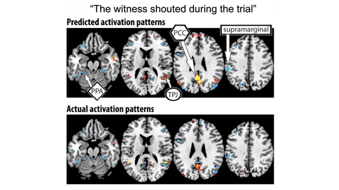
Beyond Bananas: CMU Scientists Harness “Mind Reading” Technology to Decode Complex Thoughts
By Shilo Rea
Carnegie Mellon University scientists can now use brain activation patterns to identify complex thoughts, such as, "The witness shouted during the trial."
This latest research led by CMU’s Marcel Just builds on the pioneering use of ma-chine learning algorithms with brain imaging technology to "mind read." The findings indicate that the mind’s building blocks for constructing complex thoughts are formed by the brain’s various sub-systems and are not word-based. Published in Human Brain Mapping and funded by the Intelligence Advanced Research Pro-jects Activity (IARPA), the study offers new evidence that the neural dimensions of concept representation are universal across people and languages.
"One of the big advances of the human brain was the ability to combine individual concepts into complex thoughts, to think not just of 'bananas´, but 'I like to eat bana-nas in evening with my friends,'" said Just,the D.O.Hebb University Professor of Psy-chology in the Dietrich College of Humanities and Social Sciences. "We have finally developed a way to see thoughts of that complexity in the fMRI signal. The discovery of this correspondence between thoughts and brain activation patterns tells us what the thoughts are built of."
Previous work by Just and his team showed that thoughts of familiar objects, like ba-nanas or hammers, evoke activation patterns that involve the neural systems that we use to deal with those objects. For example, how you interact with a banana involves how you hold it, how you bite it and what it looks like.
The new study demonstrates that the brain’s coding of 240 complex events, senten-ces like the shouting during the trial scenario uses an alphabet of 42 meaning com-ponents, or neurally plausible semantic features, consisting of features, like person, setting, size, social interaction and physical action. Each type of information is pro-cessed in a different brain system - which is how the brain also processes the infor-mation for objects. By measuring the activation in each brain system, the program can tell what types of thoughts are being contemplated.
For seven adult participants, the researchers used a computational model to assess how the brain activation patterns for 239 sentences corresponded to the neurally plausible semantic features that characterized each sentence.Then the program was able to decode the features of the 240th left-out sentence.They went through leaving out each of the 240 sentences in turn, in what is called cross-validation.
The model was able to predict the features of the left-out sentence, with 87 percent accuracy, despite never being exposed to its activation before. It was also able to work in the other direction, to predict the activation pattern of a previously unseen sentence, knowing only its semantic features.
"Our method overcomes the unfortunate property of fMRI to smear together the sig-nals emanating from brain events that occur close together in time,like the reading of two successive words in a sentence". Just said. "This advance makes it possible for the first time to decode thoughts containing several concepts. That’s what most human thoughts are composed of."
He added, "A next step might be to decode the general type of topic a person is thin-king about,such as geology or skateboarding.We are on the way to making a map of all the types of knowledge in the brain."
CMU’s Jing Wang and Vladimir L. Cherkassky also participated in the study.
Discovering how the brain decodes complex thoughts is one of the many brain research breakthroughs to happen at Carnegie Mellon. CMU has created some of the first cognitive tutors, helped to develop the Jeopardy-winning Watson, founded a groundbreaking doctoral program in neural computation, and is the birthplace of arti-ficial intelligence and cognitive psychology.Building on its strengths in biology, com-puter science, psychology, statistics and engineering, CMU launched BrainHub, an initiative that focuses on how the structure and activity of the brain give rise to complex behaviors. "
Yksi Jing Wang (postdoc) ja Vldimir Cherkassky (toimittaja) eivät odista muuta kuin että puuppua aioitaan syöttää myös Venäjälle ja Kiinaan.
https://www.cmu.edu/dietrich/psychology/people/core-training-faculty/just-marcel.html "

Marcel Just
D O Hebb Professor, Director CCBI , Psychology
Bio
My research uses fMRI and other technologies to uncover the structure of human thought. The fMRI studies track the brain activity that occurs during a wide range of cognitive and social thought, such as language comprehension, visual thinking, prob-lem-solving, working memory, social judgment, and multi-tasking.
One current interest is in identifying the neural basis of concept representations using fMRI in the new area of neurosemantics. In collaboration with colleagues in the School of Computer Science, we have developed experimental paradigms and ma-chine-learning techniques (multi-voxel pattern analysis) that are being applied to the study of lexical, perceptual, and social concepts (identifying the neural signature of that object and the components of the signature). We can identify the thought of a concrete object, social interaction, and digit, and we are moving on to propositions. This is leading us to a specification of how simple thoughts are neurally coded.
A second research area examines how scientific concepts are learned. As we listen to a lecture or read a textbook, neural representations of the new knowledge are being established. We hope to specify the processes by which new concepts come to be neurally represented.
Another important area of our research is in understanding the brain functioning in autism and relating it to the social and cognitive impairments that sometimes arise in the disorder. Our work has led to a new perspective,expressed as the underconnec-tivity theory of autism.This work uses fMRI and high angular resolution diffusion ima-ging (HARDI) to relate several levels of analysis: anatomical connectivity, informatio-nal (functional) connectivity, and behavioral performance. We are also starting to apply our neurosemantics (machine learning) methods to concept representations in autism.
The findings are being used to continuously develop a comprehensive theory of how brain function is related to thought, often expressed in terms of the 4CAPS computational theory.
Publications
Just, M. A., Cherkassky, V. L., Aryal, S., & Mitchell, T. M. (2010). A neurosemantic theory of concrete noun representation based on the underlying brain codes. PLoS ONE, 5, e8622.
Kassam, K.S., Markey, A.R., Cherkassky,V. L., Loewenstein, G. & Just, M. A. (2013). Identifying emotions on the basis of neural activation. PLoS ONE, 8, e66032.
Just, M. A., Keller, T. A., & Kana, R. K. (2013). A theory of autism based on frontal-posterior underconnectivity. In M. A. Just & K. A. Pelphrey (Eds.), Development and brain systems in autism (pp. 35-63). New York: Psychology Press.
Mason, R.A., Prat, C.S. & Just, M.A. (2013). Neurocognitive brain response to tran-sient impairment of Wernicke's area. Cerebral Cortex; doi: 10.1093/cercor/bhs423
PS: Miksi PSYKOLOGIT pelleilevät NEUROFYSIOLOGISTEN autismiteorioiden kanssa???!!!
***
Lack of neuron ‘pruning’ may be behind many brain-related conditions
Brain scans show that adolescents with more symptoms of certain mental health conditions, autism or ADHD have undergone less pruning than usual of synaptic connections between neurons
By Clare Wilson

Our brains prune away unwanted connections between neurons as we grow and learn MEHAU KULYK / SCIENCE PHOTO LIBRARY/Alamy
Many disparate conditions, such as depression, phobias and attention deficit hyper-activity disorder (ADHD), may have the same underlying cause: a delay in “pruning”, a process in which unneeded connections between brain cells disappear.
The finding comes from one of the largest brain-scanning studies done in adoles-cents and has been confirmed in several other data sets, including people of other ages. “We’ve been able to show that these different [conditions] are all related to a single underlying neurobiological factor,” says Barbara Sahakian at the University of Cambridge.
The results lend weight to a controversial recent idea in neuroscience that many disparate conditions share a common cause – a concept known as the p factor.
Teenagers with unhealthy lifestyles age faster than healthier peers
Until now, this was mainly based on the fact that many people have more than one such condition or may be diagnosed with different conditions at different times in their lives, as well as DNA studies finding that the same set of gene variants predispose people to multiple conditions.
Now Sahakian and her colleagues are proposing a neurobiological basis for the p factor, which they call the “neuropsychopathological (NP) factor”.
Sign up to our Health Check newsletter
Get the most essential health and fitness news in your inbox every Saturday.
The researchers did their first analysis using an existing set of brain scanning images for nearly 2000 teenagers assessed at 14, where there was also information about potentially related symptoms "
xxx
https://www.youtube.com/watch?v=jHGx7HBB-J4












.jpg)
.jpg)


.svg)
.svg)